Osteology Everywhere: Skeletal Spice

The American winter holiday season is steeped in special spices, such as nutmeg, cloves, cinnamon, and whatever the hell pumpkin spice is. I guess as part of the never-ending War on Christmas, each year this sensory and commercial immersion begins earlier and earlier. Since these have become old news, I’d pretty much forgotten about the seasonal spicecapade until just the other day. In prep for minor holiday gluttony, I was grinding fresh nutmeg when I made a startling discovery. Nutmeg is not just the fragrant fruit of the Myristica fragrans tree. No, there’s something far more sinister in this holiday staple.

download

Merely nutmeg?

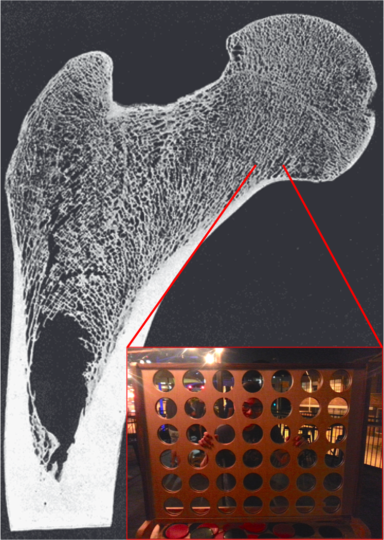
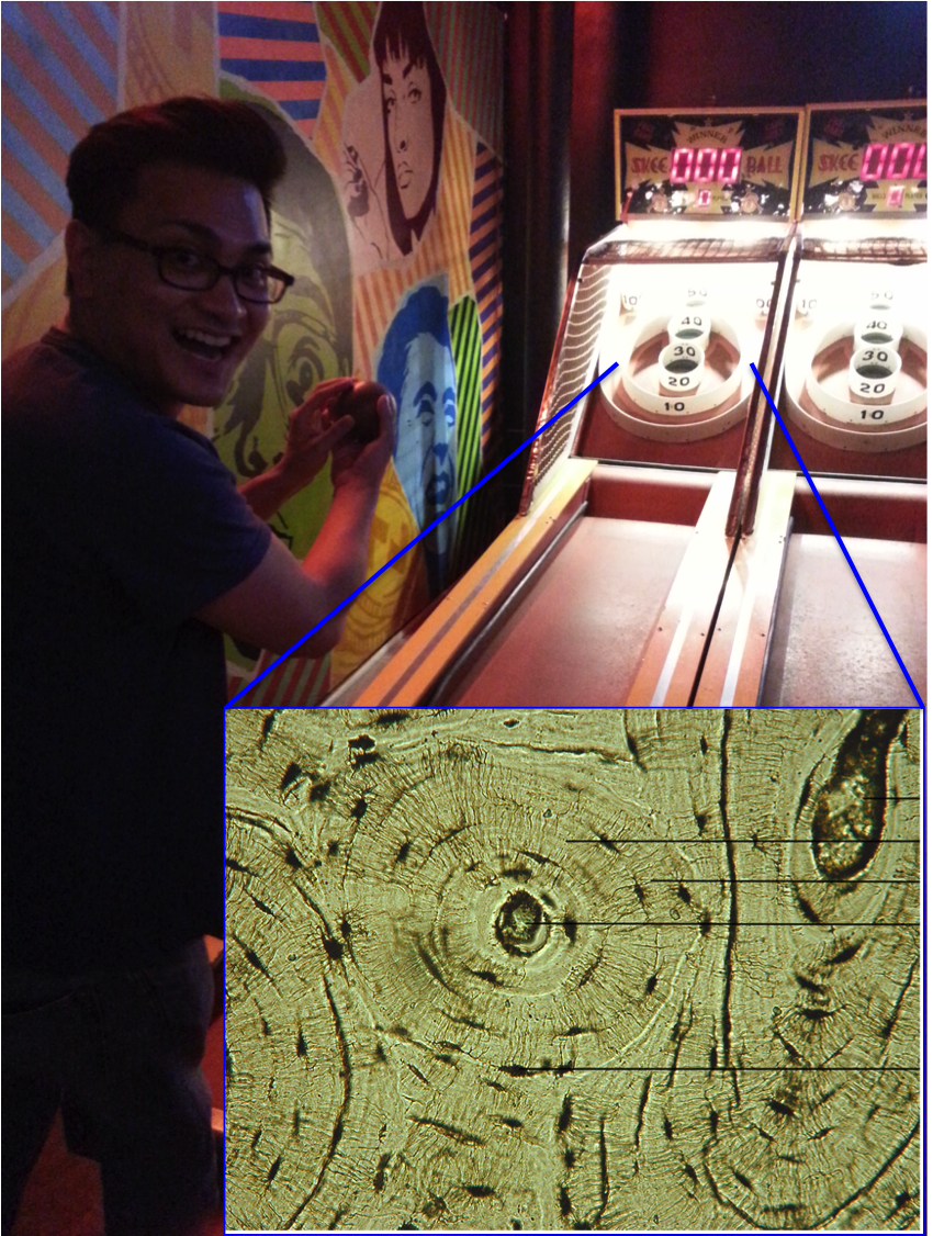
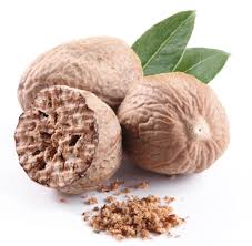
The ground section looks superficially like an unfused epiphyseal surface, whereas the rounded outer surface is more spherical. It turns out, in the most nefarious of all holiday conspiracies since the War on Christmas, nutmeg halves are nothing more than unfused femur heads! Compare with the epiphyseal surface of this Homo naledi femur head:

screen-shot-2016-11-26-at-5-36-05-pm

Nutmeg (left) and H. naledi specimen UW 101-1098 (right).

This immature H. naledi specimen was recently published (Marchi et al., in press), and the associated 3D surface scan has been available for free download on Morphosource.org for a while now. It fits onto a proximal femur fragment, UW 101-1000, also free to download from Morphosource.

screen-shot-2016-11-26-at-5-48-42-pm

Modified Fig. 11 from Marchi et al. It’s weird that only H. naledi bones were found in the Dinaledi chamber, but even weirder is the underreported presence of nutmeg.

Like most  bones in the skeleton, the femur is comprised of many separate pieces that appear and fuse together at different, fairly predictable ages. The shaft of the femur appears and turns to bone before birth, and the femur head, which forms the ball in the hip joint, usually appears within the first year of life and fuses to the femur neck in adolescence (Scheuer and Black, 2000). So we know this H. naledi individual was somewhere between 1–15ish years by human standards, probably in the latter half of this large range.

So there you have it. Osteology is everywhere – the holidays are practically a pit of bones if you keep your eyes open.

ResearchBlogging.orgREFERENCES

Marchi D, Walker CS, Wei P, Holliday TW, Churchill SE, Berger LR, & DeSilva JM (2016). The thigh and leg of Homo naledi. Journal of Human Evolution PMID: 27855981.

Scheuer L and Black S. 2000. Developmental Juvenile Osteology. New York: Elsevier Academic Press.

Osteology Everywhere: Vertebral Incidens

Try as I might, I can never escape osteology. Never. Just the other day, I was walking through my school’s expansive, boneless atrium, when these haphazardly scattered letters stopped me in my tracks:

2016-02-09 16.26.31

DЯSTUDENSN

Amidst this alphabet soup, there it was, calling out to me. Whispering. Longing….

Untitled

Ah, the dens. What is the “dens” you ask? It is a special little projection on a special little bone, the second cervical vertebra (C2). Why is it special? Well, most vertebrae look pretty similar to one another, with a body in the front being held in awkward embrace by a bony neural arch in the back.


But not the first two vertebrae, C1 and C2. No, these rebels are spinal celebrities. C1, whose rock name is “Atlas” (presumably in honor of its favorite episode of Wishbone) cradles the skull’s occipital condyles on its concave shoulders. Lacking a true body or centrum, Atlas viewed from the top resembles the gaping maw of a manta ray:

Top: Manta ray. Bottom: Atlas viewed from top, anterior is on the bottom (from Scheuer and Black, 200). A and F refer to the age at which the bony portions appear and fuse, respectively.

Top: Manta ray. Bottom: Atlas viewed from top, anterior is on the bottom (from Scheuer and Black, 2000). A and F refer to the age at which the bony portions appear and fuse, respectively.

Atlas is a jerk and so it sits right on top of C2, whose rock name is Axis (after the second album by the Jimi Hendrix Experience). More gawky and angsty than Atlas, Axis differs from the rest of the vertebrae in having an extension, the dens, which reaches skyward to boop the inside of Atlas’ maw:

Top: Axis viewed from the front. Bottom: Axis getting pwnd by Atlas. Modified from White et al. 2012.

Top: Axis viewed from the front. Bottom: Axis getting pwnd by Atlas. Modified from White et al. (2012).

The most distinctive feature of Axis, aside from its smoldering adolescent rage, is the dens (or odontoid process). If you find a bone fragment that is verily vertebral and has a perpendicular projection, you can bet good tenge you’ve got an Axis. Even a densless fragment can be distinguished from all other vertebrae by its superior articular facets, which are rather flat and face mostly superiorly.

What I thought would be a casual jaunt after class last week turned out to be a horrific reminder of the most amazing vertebrae. This must be  how Scott Williams always feels.

Osteology Everywhere: Barcade bone biology

I’ve fled the Central Asian steppe to visit my childhood home, Kansas City, Missouri.

The tortuous path from the center of Eursia to the center of the US, a mere 8500 miles since there are no direct flights. Map made by Wolfram Alpha.

The tortuous path from the center of Eursia to the center of the US, a mere 8500 miles since there are no direct flights. Map made by Wolfram Alpha.

It would be a lie to say I don’t miss life in this Midwest metropolis. Kansas City is sprawling, with diverse cultures, foods and festivities in far-flung neighborhoods. It’s always a trip to revisit the people and places of my formative years.

Of course, there are differences between now and when I was growing up. A whole new world of experiences became available to me here once I was old enough to drink (legally; this is long ago now). The bar scene itself has evolved over the past decade or so, arguably culminating in Up-Down, a grown-up video game arcade that will confusingly make you both happy and sad to have become an adult.

Be still my heart. Image credit.

I’ve never seen anything like this before. But even in this novel environment, I still couldn’t help but notice Osteology Everywhere. What appears at first glance to be an oversized Connect Four contraption . . .

Go for the bottom, go for the top.

Go for the bottom, go for the top.

. . . is in fact a closeup of trabecular bone (with my friend creepily peering through):

Section through a human proximal femur (hip joint). Note the trabecular or "spongy" bone filling the top, in comparison with the thick and dense cortical bone of the shaft in the bottom left. Image credit.

Vertical section through a human proximal femur (hip joint). Note the trabecular or “spongy” bone filling the top, in comparison with the thick and dense cortical bone of the shaft in the bottom left. Image credit.

And here, we’re not playing Skee Ball . . .

20150618_003612. . . we’re hurling wooden balls into Haversian canals and lacunae of osteons. For Science.

Cross section through cortical bone, magnified to highlight an osteon. The big hole in the center is the Haversian canal, and the smaller satellite holes are lacunae housing osteocytes.

Cross section through cortical bone, magnified to highlight an osteon. The big hole in the center is the Haversian canal, and the smaller satellite holes are lacunae housing osteocytes. Image credit.

So if you’re in the KC area, I highly recommend you check out Up-Down, where you can review osteology while also playing games and sipping a refreshing beer. Who knew learning could be so fun?

Osteology Everywhere: Head for the hills

Last week I was exploring central England with the brilliant Jess Beck, an archaeology PhD student at the University of Michigan. Both of us avid (nay, rabid) connoisseurs of everything skeletal, we espied the likes of a specific human bone in the scenic landscape of the the Cotswolds. Check out JB’s blog, Bone Broke, for her take on this geographical/geological/skeletal formation (as well as for lots of killer osteology and bioarchaeology tips and tricks). Do it now! NOW!

After you’ve checked out her site, behold this sight – what bone is lurking in the landscape?

osteourrywhere Cotswolds

As with Rorschach inkblots, probably lots of bones could be seen in this image. But what Jess & I saw was a hamate, the greener hue hewn into the hills, whose sizeable hamulus runs from the bottom right to join the rest of the carpal around the center of the image. Here’s what a real hamate looks like:

hamate

Top: Human hamate from Human Osteology (White, Black & Folkens, 2012). Bottom: rough anatomical position of the hamate in the human wrist.

The hamulus of the hamate is an attachment point for the flexor retinaculum, the band of fascia stretching across your wrist to hold your extrinsic digital flexor muscles (or rather, their tendons) in place; you could think of it as the bridge covering the carpal tunnel. Now, comparing the grassy hamulus with an actual human one, you’ll spot two important differences: first, the grassy one isn’t blunt like the humans’, but ends in a long point. Oops! Just pretend it’s rounded off. Second, the grassy hamulus is huge relative to the overall size of the bone (or valley) compared with the human form. The size of the hamulus partially reflects the size of the carpal tunnel: chimpanzees, with powerful wrists and forearms, have long hamuli.

A huge nerd, I didn’t just see any hamate in this Cotswold vale. I also immediately thought of KNM-WT 22944, an Australopithecus afarensis hamate from the 3.5 million year old site of South Turkwel in Kenya (Ward et al., 1997):

WT 22944-Ward &al 1997

From Ward et al., 1999. Sorry it’s not in the same orientation as the above image. Hamulus is the projection pointing to the bottom left corner of the “medial” image.

An absolutely and relatively massive hamulus in WT 22944 suggests whoever this bone belonged to had some powerful gripping capabilities, while a geologically younger A. afarensis hamate from Hadar (AL 333-50) had a smaller, more human-like hamulus. Maybe (some) A. afarensis were still using their arms a lot for tree-climbing, in spite of being more than capable bipeds (I’ve talked about this before here)….

One final thought: People do like the way she says, “hamate.”

Ward et al., 1999. South Turkwel: a new pliocene hominid site in Kenya. Journal of Human Evolution 36: 69-95. link

White et al., 2012. Human Osteology 3rd Edition. link