Osteology Everywhere: Bacon or first rib?

I went to a cafe today to eat breakfast and get some work done. Write, write, write. It’s important to be properly nourished to ensure maximal productivity.

The Ron Swanson diet.

The Ron Swanson diet.

But I was aghast to behold the food they placed before me:

More bacon, please.

What on earth is this?

First of all, this is not a sufficient amount of bacon.

ljQEkGT

Secondably, this bacon is a spitting image of a first rib:

First ribs, from left to right: Human, chimpanzee, bacon. First two images from eSkeletons.org.

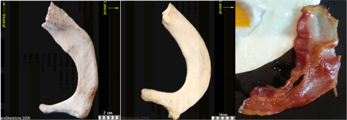
First ribs from the right side of the body, viewed from the top. From left to right: Human, chimpanzee, bacon. First two images from eSkeletons.org.

At the top of the ribcage, just beneath the clavicle and subclavian artery and vein, the first rib is much shorter and flatter than the rest of the ribs. As Jess Beck at Bone Broke points out, “The first and second rib give something of an awkward ‘slow song at a middle-school dance’ kind of a hug, while the lower ribs provide a more comfortable and self-assured embrace.” I mean, just lookit how sheepishly the bacon dances with the eggs in the first picture, it has ‘middle-school dance’ written all over it.

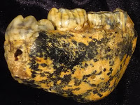
But the bacon is not totally identical to the human and chimpanzee counterparts. It’s missing their anteromedially sweeping arc, and the distal portion reaching out to the egg is fairly straight. This suggests we’re probably missing much of the original distal end. Posteriorly or dorsally (toward the bottom in the pic), it also appears to be missing much of the lateral portion including the vertebral facet. In this regard, this bacon rib looks a lot like the first rib of Homo naledi:

Full stack of ribs. From left to right: Human, bacon, Homo naledi, Dmanisi Homo erectus, Australopithecus sediba (x2), Australopithecus afarensis specimen "Lucy," Ardipithecus ramidus, and chimpanzee. Images not to scale except Lucy and Ardi.

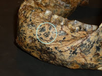
Full stack of ribs. Left to right: Human, bacon, Homo naledi, Dmanisi Homo erectus, Australopithecus sediba (x2), Australopithecus afarensis specimen “Lucy,” Ardipithecus ramidus, and chimpanzee. Images not to scale except Lucy and Ardi. Image credits given below.

It is hard to make good homologous comparisons among these fossils and bacon, since so many are so incomplete. But it looks like the hominins are relatively longer (front to back, or dorsoventrally) compared to the chimpanzee. That is, oriented along the rib “neck,” the ventral/distal end projects far more medially beyond the proximal vertebral facet in the chimp, while in the hominins the two ends are more flush.  Ardi is really incomplete and so very hard to orient, but it may be more like the chimp (I think it needs to be rotated to the right more, to make the lateral edge more vertical like all the other specimens).

It will be interesting to see what my colleagues working on the Homo naledi thorax have to say about rib shapes and their functional importance, hopefully not too long from now.

Anyway, I really wish I had more bacon.

Fossil rib sources
ResearchBlogging.orgDmanisi Homo erectus: Lordkipanidze D, Jashashvili T, Vekua A, Ponce de León MS, Zollikofer CP, Rightmire GP, Pontzer H, Ferring R, Oms O, Tappen M, Bukhsianidze M, Agusti J, Kahlke R, Kiladze G, Martinez-Navarro B, Mouskhelishvili A, Nioradze M, & Rook L (2007). Postcranial evidence from early Homo from Dmanisi, Georgia. Nature, 449 (7160), 305-10 PMID: 17882214

Australopithecus sediba: Schmid P, Churchill SE, Nalla S, Weissen E, Carlson KJ, de Ruiter DJ, & Berger LR (2013). Mosaic morphology in the thorax of Australopithecus sediba. Science, 340 (6129) PMID: 23580537

Homo naledi: Morphosource.

Australopithecus afarensis and Ardipithecus ramidus: White TD, Asfaw B, Beyene Y, Haile-Selassie Y, Lovejoy CO, Suwa G, & WoldeGabriel G (2009). Ardipithecus ramidus and the paleobiology of early hominids. Science, 326 (5949), 75-86 PMID: 19810190

These new fossils are intriguing as hell

Some big changes here at Lawnchair Anthropology. I just successfully defended my dissertation (Mandibular Growth in Australopithecus robustus, more info on that to come), and moved to Kazakhstan to begin my new job in the School of Humanities and Social Sciences at Nazarbayev University. I landed in Astana about 22 hours ago, so I should be asleep, battling (or succumbing to) jetlag, but some friends have pointed me to newly published early Homo fossils from Kenya, dating to between 1.9-1.6 million years ago (Leakey et al., 2012). See Adam Van Arsdale’s blog, the Pleistocene Scene, for great historical background and perspective on these new fossils.

Now, one of the themes of my dissertation is that there is lots of interesting information to be gleaned from fossils that we’ve known about for a long time (many of the A. robustus mandibles featured in my research have been known for decades). But dammit if some of these much more recently discovered fossils point to tantalizing variation in hominids just later than 2 million years ago (note I’m careful to say “variation” rather than “diversity”). In light if this variation, Adam discusses the similarities between one of these Kenyan fossils (KNM-ER 60000) and the large mandible from Dmanisi, which was discovered in only in the year 2000 (Gabunia et al., 2002).

Piggy-backing off Adam, I’d like to point out similarities between another of the new fossils, the KNM-ER 62000 face of a juvenile, and the recently discovered A. sediba juvenile face (Berger et al., 2010). These two fossils are at the same stage of dental development, so they’re roughly at the same stage of life. They are close in geological age, but A. sediba is from South Africa. Below are figures of A. sediba (left) and the ER 62000 face (right). The pics should be to scale, modified from the original publications. (sorry I couldn’t remove the background from the top left one)

What do you think? Pretty different, right? WRONG! Below I’ve superimposed the ER 62000 face onto A. sediba (slightly recolored and transparented for contrast). Remember that these are to scale.

In front view (left), the ER 62000 face is almost identical to A. sediba, right down to the positions of the teeth. THIS DOES NOT MEAN THAT I THINK THESE TWO FOSSILS REPRESENT THE SAME SPECIES. In side view, however, some differences do become apparent. Notably, the front of the A. sediba maxilla projects a bit further forward than ER 62000, and the nasal and orbital anatomy are also fairly different. THIS DOES NOT MEAN THAT I THINK THESE ARE DIFFERENT SPECIES. (although I would be surprised if these fossils turned out to be the same animal)

Leakey et al. liken these new Kenyan fossils to the cranium KNM-ER 1470, from the same region and at 1.9 million years old. But what’s weird to me is that ER 1470 actually looks a bit more like the juvenile A. sediba in the side view (as reconstucted; the face and braincase of ER 1470 are actually separated, leaving it unclear just how the two parts fit together). Here are all three specimens, to scale:

From left to right: ER 62000, A. sediba, ER 1470

Now, the ER 1470 comparison isn’t really fair – ER 1470 is an adult and it is much larger: the bottom of ER 1470’s eye socket is about the same height as the top of A. sediba‘s. The size difference is probably the main reason why its face below the nose sticks out as much as A. sediba‘s, even though the latter is smaller. (I should note, too, that the adult A. sediba mandible is superficially very similar in gonial and ramus anatomy to another of the recently published Kenyan specimens, ER 60000).

The point of all these comparisons is not to say whether these fossils are the same species, but rather to point out that there are actually striking similarities between fragmentary fossils, and it’s not clear what exactly these similarities (or differences, for that matter) mean. Maybe my eye was drawn to the ER 62000-A. sediba comparison not because of any evolutionary relationship, but because these fossils are in similar stages of growth and development – if it weren’t waaaaay past my bedtime I’d love to compare these fossils with other similarly-aged fossils (like D2700 from Dmanisi and KNM-WT 15000, also from Kenya).

All of these fossils (except ER 1470) were discovered in the past few years. I’ve said it before and I’ll repeat it now: this is a great time to study paleoanthropology.

ResearchBlogging.orgRead more NOW
Berger L, de Ruiter DJ, Churchill SE, Schmid P, Carlson KJ, Dirks PHGM, and Kibii JM. 2010. Australopithecus sediba: A New Species of Homo-like Australopith from South Africa. Science 328: 195 – 204.

L. Gabounia, M.-A. de Lumley, A. Vekua, D. Lordkipanidze, and H. Lumley. 2002. Découverte d’un nouvel hominidé à Dmanissi (Transcaucasie, Géorgie). Comptes Rendus Palevol 1(4):243-253

Meave G. Leakey, Fred Spoor, M. Christopher Dean, Craig S. Feibel, Susan C. Antón, Christopher Kiarie, & Louise N. Leakey (2012). New fossils from Koobi Fora in northern Kenya confirm taxonomic diversity in early Homo Nature, 408, 201-204 DOI: 10.1038/nature11322

Pictures worth thousands of words and dollars

ResearchBlogging.orgLooking into subdural empyema, which is a meningeal infection you don’t want, I stumbled upon a study from the roaring 1970s – the glorious Nixon-Ford-Carter years – using computerized axial tomography (hence, CAT scan) to visualize lesions within the skull (Claveria et al. 1976). Nowadays people refer to various similar scanning techniques simply as “CT” (for computed tomography, though this is not exactly the same as magnetic resonance imaging, MRI).

It’s pretty amazing how medical imaging has advanced in the 35 years since this study. For example, to the right is a CAT scan from Claveria et al. (1976, Fig. 4). These are transverse images (“slices”) through the brain case, the top of the images corresponding to the front of the face. You can discern the low-density (darker) brain from the higher density (lighter) bone – the sphenoid lesser wings and dorsum sellae, and petrous pyramids of the temporal bones are especially prominent in the top left image. In the bottom two images you can see a large, round abscess in the middle cranial fossa. Whoa.

What makes this medical imaging technique so great is that it allows a view inside of things without having to dissect into them. Of course, the downside is that it relies on radiation, so ethically you can’t be so cavalier as to CT scan just any living thing. If I’d been alive in 1976, CAT scanning would’ve blown my mind. Still, the image quality isn’t super great here, there’s not good resolution between materials of different densities, hence the grainy images.

But since then, some really smart people have been hard at work to come up with new ways to get better resolution from computerized tomography scans, and the results are pretty amazing. To the left is a slice from a synchrotron CT scan of the MH1 Australopithecus sediba skull (Carlson et al. 2011, Supporting on line material, Fig. S10). You’re basically seeing the fossil face-to-face … if someone had cut of the first few centimeters of the fossil’s face. Just like the movie Face Off.

Quite a difference from the image above. Here, we can distinguish fossilized bone from the rocky matrix filling in the orbit, brain case and sinuses. Synchrotron even distinguishes molar tooth enamel from the underlying dentin (see the square). The post-mortem distortion to the (camera right) orbit is clear. It also looks as though the hard palate is thick and filled with trabecular bone, as is characteristic of robust Australopithecus (McCollum 1999). Interesting…

Even more remarkable, the actual histological structure of bone can be imaged with synchrotron imaging. Mature cortical bone is comprised of these small osteons (or Haversian systems), that house bone cells and transmit blood vessels to help keep bone alive and healthy. Osteons are very tiny, submillimetric. To the right is a 3D reconstruction of an osteon and blood vessels, from synchrotron images (Cooper et al. 2011). The scale bar in the bottom right is 250 micrometers. MICROmeters! Note the scan can distinguish the Haversian canal (red part in B-C) from vessels (white part in B). Insane!

Not only has image quality improved over the past few decades, but CT scanning is being applied outside the field of medicine for which it was developed; it’s becoming quite popular in anthropology. What I’d like to do, personally, with such imaging is see if it can be used to study bone morphogenesis – if it can be used to distinguish bone deposition vs. resorption, and to see how these growth fields are distributed across a bone during ontogeny. This could allow the study the proximate, cellular causes of skeletal form, how this form arises through growth and development. If it could be applied to fossils, then we could potentially even see how these growth fields are altered over the course of evolution: how form evolves.

References
Carlson KJ, Stout D, Jashashvili T, de Ruiter DJ, Tafforeau P, Carlson K, & Berger LR (2011). The endocast of MH1, Australopithecus sediba. Science (New York, N.Y.), 333 (6048), 1402-7 PMID: 21903804

Claveria, L., Boulay, G., & Moseley, I. (1976). Intracranial infections: Investigation by computerized axial tomography Neuroradiology, 12 (2), 59-71 DOI: 10.1007/BF00333121

Cooper, D., Erickson, B., Peele, A., Hannah, K., Thomas, C., & Clement, J. (2011). Visualization of 3D osteon morphology by synchrotron radiation micro-CT Journal of Anatomy, 219 (4), 481-489 DOI: 10.1111/j.1469-7580.2011.01398.x

McCollum, M. (1999). The Robust Australopithecine Face: A Morphogenetic Perspective Science, 284 (5412), 301-305 DOI: 10.1126/science.284.5412.301

[insert clever quip about australopithecus hips]

A week and a half ago, Kibii and colleagues (2011) published reconstructions and re-analyses of two hips belonging to the 1.98 million-year old Australopithecus sediba. As with many fossil discoveries, these additions to the fossil record raise more questions than they answer. Unless the question was, “did A. sediba have a pelvis?” It did. Here’s a good summary from the paper itself:

Thus, Au. sediba is australopith-like in having a long superior pubic ramus and an anteriorly positioned and indistinctly developed iliac pillar…[and] Homo-like in having vertically oriented and sigmoid shaped iliac blades, more robust ilia, and a narrow tuberoacetabular sulcus…and the pubic body is upwardly rotated as in Homo. (p. 1410, emphases mine)

So far as I can tell, the main way the hips are ‘advanced’ toward a more human-like condition is that the iliac blades are more upright and sweep forward more than in earlier known hominid hips. Here’s the figure 2 from the paper (more sweet pics of the fossils are available here). NB that in both A. sediba hips much of the upper portions of the iliac blades are missing (reconstructed in white; this region is missing in lots of fossils), so it’s possible they were more flaring like the australopith in the center photo.

The authors’ bottom-line, take-home point is that the A. sediba pelvis has features traditionally associated with large-brained Homo – but belonged to a small-brained species (based solely on the ~430 cc MH1 endocast). They argue that this means that many of these unique pelvic features did not evolve in the context of birthing large-brained babies, as has often been thought. They state that these features are thus “most parsimoniously attributed to altered biomechanical demands on the pelvis in locomotion,” and suggest that this hypothetical locomotion was mostly bipedalism but with a good degree of climbing. Maybe, maybe not. This interpretation is consistent with the analysis of the A. sediba foot/ankle (Zipfel et al. 2011).

The weird mix of ancient (australopith-like) and newer (Homo-like) pelvic features in A. sediba really raises the question of how australopithecines moved around. More intriguing is that the A. sediba pelvis has different Homo-like features than the ~1 million year old Busidima pelvis (Simpson et al. 2008), which has been attributed to Homo erectus (largely in aspects of the iliac blades). This raises the question of whether A. sediba is really pertinent to the origins of the genus Homo, and whether the Busidima pelvis belongs to Homo erectus or a late-surviving robust australopithecus (e.g. boisei, Ruff 2010).

Also interesting is that the subpubic angle (in the pic above, the upside-down “V” created by the pubic bones just above the red labels) is pretty low in MH2. This is curious because modern human males and females differ in how large this angle is – females tend to have a large angle which contributes to an enlarged birth canal, whereas males have a low angle like MH2. But MH2 is considered female based on skeletal and dental size. This raises the additional questions of whether human-like sexual dimorphism had not evolved in hominids prior to 1.9 million years ago, and whether the sex of MH2 was accurately described.

Finally, though the authors did a great job comparing this pelvis with those from other hominids, I think a major, more comprehensive comparative review of hominid pelves is in order. How does the older A. afarensis hip from Woranso (Haile-Selassie et al. 2010) inform australopithecine pelvic evolution? What about the possibly-contemporary-maybe-later hip from the nearby site of Drimolen (Gommery et al. 2002)? Given the subadult status of the MH1 individual, it would be interesting to compare with the WT 15000 Homo erectus fossils, or A. africanus subadults from Makapansgat, to examine the evolution of pelvic growth.

ResearchBlogging.org

Lots of interesting questions arise from these fascinating new fossils. “The more you know,” right?

References
Gommery, D. (2002). Description d’un bassin fragmentaire de Paranthropus robustus du site Plio-Pléistocène de Drimolen (Afrique du Sud)A fragmentary pelvis of Paranthropus robustus of the Plio-Pleistocene site of Drimolen (Republic of South Africa) Geobios, 35 (2), 265-281 DOI: 10.1016/S0016-6995(02)00022-0

Haile-Selassie Y, Latimer BM, Alene M, Deino AL, Gibert L, Melillo SM, Saylor BZ, Scott GR, & Lovejoy CO (2010). An early Australopithecus afarensis postcranium from Woranso-Mille, Ethiopia. Proceedings of the National Academy of Sciences of the United States of America, 107 (27), 12121-6 PMID: 20566837

Kibii, J., Churchill, S., Schmid, P., Carlson, K., Reed, N., de Ruiter, D., & Berger, L. (2011). A Partial Pelvis of Australopithecus sediba Science, 333 (6048), 1407-1411 DOI: 10.1126/science.1202521

Ruff, C. (2010). Body size and body shape in early hominins – implications of the Gona Pelvis Journal of Human Evolution, 58 (2), 166-178 DOI: 10.1016/j.jhevol.2009.10.003

Simpson, S., Quade, J., Levin, N., Butler, R., Dupont-Nivet, G., Everett, M., & Semaw, S. (2008). A Female Homo erectus Pelvis from Gona, Ethiopia Science, 322 (5904), 1089-1092 DOI: 10.1126/science.1163592

Zipfel, B., DeSilva, J., Kidd, R., Carlson, K., Churchill, S., & Berger, L. (2011). The Foot and Ankle of Australopithecus sediba Science, 333 (6048), 1417-1420 DOI: 10.1126/science.1202703

New Australopithecus sediba analyses

A slew of papers analyzing the brain, hands, feet, and pelvis of Australopithecus sediba were just published in the journal Science. I have not had a chance to read them yet – nor will I for a few days as I’m in a wedding Saturday [not mine 😦 ] and the partying starts in a few hours. So I’m afraid I won’t be able to report on and interpret these on the blog for a while. Please stay tuned!

CT reconstruction, from Science (follow link above).
The exact same thing happened to me 2 years ago when the Ardipithecus ramidus skeleton was (finally) published. I remember waiting in the Detroit airport to board a flight to St. Louis to begin my platonic soul-mate’s bachelor party, and I get a flurry of emails on my phone announcing the skeleton in the 15 year old closet.
So media beware – whenever I’m in a wedding, badass new fossil studies will be published. 

eFfing Fossil Friday – Renaissance and Designer Fossils

Sorry I’m a bit late on this one, and that I’ve fallen behind on keeping the blog updated. I’ve been scrambling to make all the observations on, and collect all the data from, these Australopithecus robustus mandibles in a short time. As my advisor likes to remind me, everything always takes 3x longer than you initially anticipate, and this is certainly true of my work here. Yesterday (the actual Fossil Friday), in fact, I probably spent only 30 min with these fossils. Instead, I accompanied Lee Berger and John Hawks on a trip to Malapa – the site that recently yielded fossils of the mysterious Australopithecus sediba – and other sites in the area. To get there, I rented a car and drove on the wrong side of the road for the first time – it was a trippy trip, every time I got in the car I reached to my left for a phantom seat belt, and kept searching for the gear-shift my mind thought was in the door. Nuttiness.
Anyway, I have two thoughts for this edition of eFfing Fossil Friday. First point, related to the great tour from Dr. Berger, is that a ton of hominid fossils are lying in wait for us to re-expose them to the light of day. In South Africa, the classic Plio-Pleistocene sites have been Makapansgat (A. africanus), Sterkfontein (A. africanus) and Swartkrans (A. robustus and early Homo). These sites have variously been worked since the early 20th century. Since then, a number of other hominid-bearing sites – largely in the same area as Sterkfontein and Swartkrans – have been discovered: Gladysvale, Gondolin, Drimolen, and most recently Malapa. Yet still a metric-tonne of work is still being done on the more classic sites (except maybe Makapansgat?).
View of the valley, Malapa is somewhere in the background, I think the green patch of trees near the center, just before the big hill-shadow (?).
But these sites are just the tip of a fossiliferous iceberg. A few years ago when I was working here I accompanied some other researchers on a survey for more fossil sites in the area. What I learned then is that if you look across the Sterkfontein valley in the winter, the dessicated grassland is pimpled with the occasional patch of green trees – these small verdant isles are the tells of underlying cave systems (the caves contain water that plants will cut throats for). What was driven home yesterday at Malapa and other sites Dr. Berger showed us, is that these caves are all over the place, many fossil treasure-troves. What’s more, the A. sediba discovery (and the massive hominid molars from Gondolin) points to the idea that we are only beginning to understand what hominid life was like in the past. There is a rich prehistory still waiting to be discovered in South Africa, and undoubtedly also the rest of the African continent. Human paleontological work is far from exhausted. Let us usher in a Renaissance of field Paleoanthropology!
My next thought is that the process of fossilization can make the fossil-memories of past life quite beautiful. Now, in life the enamel of teeth is white-ish (yellow/brown is also not uncommon), and bone is this off-white/yellowish color. But during the process of fossilization, the original minerals used to make the bone (and less commonly teeth) are replaced by those in the surrounding soil. Often these minerals gussy up the fossils in neat new ways – manganese for example tends to make bone/tooth black.


Check out SK 61, an infant/child Australopithecus robustus. After fossilization, this thing takes on a designer, tortoise-shell coloration (left, above). SK 12, an older adult A. robustus (right, above), is another good example: some subterranean joker has drawn a smiley face beneath his left premolar (circled). So while we are often left with a meager fossil record, at least the fragments we get are voluptuously variegated.

Holy Effing Crap II: Australopithecus from Malapa

Lee Berger and colleagues report in Science today on 2 incredibly well preserved skeletons – including perhaps the best-preserved hominid skull in South Africa, in some ways as good as or better than Sts 5 (Australopithecus africanus). The specimens come from a site called Malapa in South Africa, dating to around 1.9 – 1.7 million years ago. The authors argue that it is so unique in its features that it warrants a new species – Australopithecus sediba – linking the earlier A. africanus with later Homo habilis. Is it really a new species? In my personal opinion, there’s not much distinguishing it from A. africanus.
The amazingly preserved skull is of a subadult, maybe 11 years old. The highly angled root of the zygomatic, positioned just above the M1 alveolus is classic A. africanus. It really reminds me of Sts 17, or possibly Sts 52 in the lower face. The prognathism is modest and lacks the anterior nasal pillars which tend to be fairly pronounced in A. africanus; in this regard it is quite comparable to specimens like TM 1512. Like Sts 52, it has multiple infraorbital foramina. The cranial capacity is estimated at 420 cubic centimeters (cc), which is pretty small, but within the estimated A. africanus range of variation. The authors say that the relatively low position of the temporal lines, spaced far apart from the sagittal suture, and the fact that the zygomatic arches do not flare to the sides, are features more like Homo than like A. africanus. But the specimen is only 11 years old; while the brain is finished growing, the face and chewing muscles probably aren’t. So if this were a fully adult specimen, I’m sure both of these features would come to look more like A. africanus.
The upper and lower first and second molars increase in size posteriorly, and have a distinct protostylid (enamel shelf along the side of one of the cusps) which has a very high frequency in A. africanus. The upper molars, while not totally complete, preserve something that I’ve noticed and I’m sure is in the literature, that the M1 is fairly small and squared compared to the generally larger and not-quite-as-square M2. In a few words, then, the skull seems to fit comfortably in the range of A. africanus variation.
Perhaps the least A. africanus-like aspect of the skull is the supraorbital torus. The supraorbital, or brow, is generally a modestly expressed in most africanus specimens that preserve it. The Malapa specimen is much more similar, to my eye, to later Homo in its projection and arching over the eyes. What could this mean? Moss and Young’s (1960) functional matrix model of looking at the cranium views the supraorbital as a function of the relative position of the brain to the orbits. Perhaps the spatial relationship between the vault and the face which becomes characteristic of later homo becomes established in earlier in the lineage. Other than the supraorbital, this specimen seems purely A. africanus to me. In all, the contour of the vault may not be too different from younger Dmanisi specimens like D2700 or 2280; that Malapa lacks the occiput gives an artificially short front-to-back look to the specimen. The face, however, is totally A. africanus.
Perhaps one of the most striking images is the lateral view. This photo looks strikingly similar to a subadult chimpanzee, albeit with a taller face and less prognathic snout. Maybe I’ve just seen a subadult ape before that this thing reminds me of.
So, this is an immensely exciting set of fossils. Is there a new species of Australopithecus? I wouldn’t bet my life on it. If you go with the widely held idea, that A. africanus or something like it was ancestral to later A. robustus on the one hand, and our Homo ancestors on the other, this thing would fall on the Homo side of that split. So in this case, since we’re not seeing anagenetic evolution – evolutionary change within a lineage – but rather branching, how do you name this thing? It might be slightly more derived toward a Homo than either its “pure” africanus ancestors and A. robustus evolutionary cousins, does this make it Homo? The issue is that adaptively its morphology doesn’t seem to be different from A. africanus, which would argue against the generic distinction. But if its later ancestors become H. habilis and nothing else, then I suppose this would make it a “chronospecies” of H. habilis. So maybe we should call this thing H. habilis? I think most people would argue with this simply on the brain size issue. And the brain is way smaller than any proclaimed Homo specimen.
Taxonomically, this will be a tough call.
References
Berger L, de Ruiter DJ, Churchill SE, Schmid P, Carlson KJ, Dirks PHGM, and Kibii JM. 2010. Australopithecus sediba: A New Species of Homo-like Australopith from South Africa. Science 328: 195 – 204
Moss ML and Young R. 1960. A functional approach to craniology. American Journal of Physical Anthropology 18: 281 – 292