The most complete Australopithecus skeleton

StW 573, a hominin skeleton more palatably nicknamed “Little Foot,” made its big debut last week:

800 (3)

Ron Clarke showcases the lovingly-excavated skeleton (Photo credit: AP/Themba Hadebe)

The skeleton is remarkable in that it is the most complete australopithecine individual ever discovered, and is among the most complete in the entire hominin fossil record. Below I’ve compared it to the most complete Australopithecus afarensis (KSD-VP-1/1 and AL 288-1), A. africanus (StW 431 and Sts 14), and A. sediba (MH1-2); the Dikika infant would be a neat comparison, too, but I don’t know of any photos of its bones nicely laid out. The other skeletons are practically naked (or dismembered) compared to Little Foot.

Australopithecus skeletons.png

Little Foot (red) compared with other australopithecine skeletons. Images not to scale! (Photo credit: The Internet!)

Beyond it’s completeness, the other parts of story of Little Foot are equally fascinating – from its discovery based on already-known fragments to the possibility that it is older than “Lucy” (AL-288). Ron Clarke has painstakingly and I’d say very successfully removed the skeleton from the hard breccia in which the fossils were encased. Having spent the better part of the past two decades with the skeleton, he has argued that Little Foot represents a second hominin species at Sterkfontein, Australopithecus prometheus (Clarke 2013), the species to which hominin fossils at Makapansgat were originally attributed (Dart 1948). With the unveiling of the skeleton, I’d guess that in the coming years we’ll see renewed investigations into the number of species at Sterkfontein, and the general comparison between hominins from there and Makapansgat.

From pictures in the media releases, we can see a few things that weren’t known from previous publications. I’ll outline a few here, but emphasize that these are only  superficial observations and will need to be borne out by further research.

800 (5)

“EXPELLIARMUS”

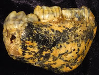
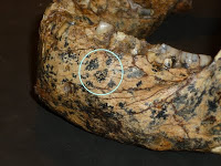
At the top of the trunk, the cervical vertebra seems to have a fairly wide spinal canal, a human-like ‘bulging’ which Meyer and Hausler (2015) suggest might reflect innervation of highly manipulative hominin hands.

Stw 573 thorax

Close up of the skull and upper trunk of StW 573, highlighting the cervical vertebral canal (white arrow) and first rib (orange arrow). Original photo credit: AP//Themba Hadebe.

In addition, the first rib may be relatively long front-to-back (as opposed to wide side-to-side), possibly indicating a more barrel-like chest than in other early hominins; the angle of the photo and the clear break between the proximal and distal portions, however, makes this unclear.

Screen Shot 2017-12-11 at 9.21.13 AM

Hominin first ribs/bacon, with StW 573 on the far right. Not to scale! Modified from this post.

The distal forelimb (i.e., radius and ulna) are not as elongated as in apes, but the femur is not as elongated as in the genus Homo. From the pictures, the femur neck appears short like in humans, not as elongated as is characteristic for australopiths and early Homo.

Limb comparison Stw 573

Limb proportion comparison. Humerus (top row), radius & ulna (middle), and femur (bottom). Image modified from Asfaw et al. (1999). StW 573 scaled to same humerus length as the human. Note also that all bones are from the right except the StW 573 upper limb.

The apparently short femur neck, similar to humans, contrasts with the wide, flaring ilium of the pelvis. This appears fairly flat, short and wide (Australopithecus af) compared to modern humans’ more strongly curved ilium. But this inference is just from a picture and it’s likely that the fossil needs a bit of reconstruction to uncover the true anatomy.

Screen Shot 2017-12-08 at 3.28.27 PM

StW 573 pelvis (left) compared with Sts 14 (A. africanus, middle) and SH pelvis 1 (archaic Homo, right). Sts 14 modified from Berge & Goularis (2010) and SH pelvis 1 from Bonmati et al. (2010).

I’d like to emphasize that these are just first impressions based on press release photos, and actual analysis of the skeleton are necessary to tell if these impressions are correct. As could be expected, the skeleton as a whole looks typically australopithecine, although the short femur neck may be a bit different. As 2017 draws to a close, let’s hope 2018 sees the testing of these predictions.

References

Asfaw B. et al. 1999. Australopithecus garhi: a new species of early hominid from Ethiopia. Science 284: 629-635.

Berge C and Goularis D. 2010. A new reconstruction of Sts 14 pelvis (Australopithecus africanus) from computed tomography and three-dimensional modeling techniques. Journal of Human Evolution 58: 262-272.

Bonmati A. et al. 2010.Middle Pleistocene lower back and pelvis from an aged human individual from the Sima de los Huesos site, Spain. Proceedings of the National Academy of Sciences 107: 18386-18391.

Clarke RJ. 2013. Australopithecus from Sterkfontein Caves, South Africa. In The Paleobiology of Australopithecus, Reed et al., eds. Dordrecht: Springer Science+Business.

Dart R. 1948. The Makapansgat proto-human Australopithecus prometheusAmerican Journal of Physical Anthropology 6: 259-284.

Meyer M. and Hausler M. 2015. Spinal cord evolution in early Homo. Journal of Human Evolution 88: 43-53.

Osteology Everywhere: Aerial Ossicles

Last month I was flying down to New Orleans for the AAPA conference. I was excited to try authentic beignets & sazeracs, present new research, and catch up with colleagues. Midway through the flight I glanced out the window, not expecting to see much. But lo!

twilight

Thankfully there wasn’t something on the wing. But there was something strange out there in the sparkle of sprawling city lights:

IMG_20170419_084210_479

What’s that I spy outside the city center?

A bit outside of the main jumble of street lamps appears to be a concentration of light superficially similar to an incus, one of the three auditory ossicles of the middle ear:

nightmare

Left: An osteologist’s nightmare at 20,000 feet. Right: Ear ossicles from White et al. (2012).

As a good mammal, there are three small bones inside your middle ear. These are fully formed at birth, and help transfer and amplify sound vibrations from your eardrum to your inner ear. It’s nuts. What’s even more nuts is that paleontologists and anatomists have figured out that the tiny, internal incus and malleus of mammals evolved from larger, external pieces of the jaws of our pre-mammalian ancestors. INSANITY!

anatomy-of-the-inner-ear-chart2b1

Cross section of a right ear, viewed from the front. Image credit.

Being so tiny, it’s not surprising that auditory ossicles are not often recovered from skeletal remains, and are pretty rare in the human fossil record. Nevertheless, some are known and their comparison with humans’ ossicles is pretty interesting. The oldest incudes (yes, the plural of incus is incudes) I know of are from SK 848 and SKW 18Australopithecus robustus fossils from Swartkrans in South Africa (Rak and Clarke, 1979; Quam et al., 2013). SK 848 is on the left in the set of images below:

Ossicles

Incus bones in three different views of SK 848, human chimpanzee, gorilla, sock puppet (left to right). Modified from Rak and Clarke, 1979.

SK 848 to differs from humans and African apes in looking more like a screaming sock puppet with a horn on the back of its head. Additional ossicles are known from South African australopithecines, including the older A. africanus from Sterkfontein (Quam et al., 2013). Interestingly, malleus of these hominins is very similar to that of humans, and Quam et al. (2013) think this ossicle may be one of the first bones in the entire skeleton to take on a human-like configuration during hominin evolution. Functionally, this may mean that the frequency range to which human ears are adapted may have appeared pretty early in our lineage as well (Quam et al., 2015).

Who’d’ve thunk we’d learn so much just from looking out an airplane window?

anthropology
ResearchBlogging.orgRead more!

Quam, R., de Ruiter, D., Masali, M., Arsuaga, J., Martinez, I., & Moggi-Cecchi, J. (2013). Early hominin auditory ossicles from South Africa Proceedings of the National Academy of Sciences, 110 (22), 8847-8851 DOI: 10.1073/pnas.1303375110

Quam, R., Martinez, I., Rosa, M., Bonmati, A., Lorenzo, C., de Ruiter, D., Moggi-Cecchi, J., Conde Valverde, M., Jarabo, P., Menter, C., Thackeray, J., & Arsuaga, J. (2015). Early hominin auditory capacities Science Advances, 1 (8) DOI: 10.1126/sciadv.1500355

Rak Y, & Clarke RJ (1979). Ear ossicle of australopithecus robustus. Nature, 279 (5708), 62-3 PMID: 377094

Osteology Everywhere: Skeletal Spice

The American winter holiday season is steeped in special spices, such as nutmeg, cloves, cinnamon, and whatever the hell pumpkin spice is. I guess as part of the never-ending War on Christmas, each year this sensory and commercial immersion begins earlier and earlier. Since these have become old news, I’d pretty much forgotten about the seasonal spicecapade until just the other day. In prep for minor holiday gluttony, I was grinding fresh nutmeg when I made a startling discovery. Nutmeg is not just the fragrant fruit of the Myristica fragrans tree. No, there’s something far more sinister in this holiday staple.

download

Merely nutmeg?

The ground section looks superficially like an unfused epiphyseal surface, whereas the rounded outer surface is more spherical. It turns out, in the most nefarious of all holiday conspiracies since the War on Christmas, nutmeg halves are nothing more than unfused femur heads! Compare with the epiphyseal surface of this Homo naledi femur head:

screen-shot-2016-11-26-at-5-36-05-pm

Nutmeg (left) and H. naledi specimen UW 101-1098 (right).

This immature H. naledi specimen was recently published (Marchi et al., in press), and the associated 3D surface scan has been available for free download on Morphosource.org for a while now. It fits onto a proximal femur fragment, UW 101-1000, also free to download from Morphosource.

screen-shot-2016-11-26-at-5-48-42-pm

Modified Fig. 11 from Marchi et al. It’s weird that only H. naledi bones were found in the Dinaledi chamber, but even weirder is the underreported presence of nutmeg.

Like most  bones in the skeleton, the femur is comprised of many separate pieces that appear and fuse together at different, fairly predictable ages. The shaft of the femur appears and turns to bone before birth, and the femur head, which forms the ball in the hip joint, usually appears within the first year of life and fuses to the femur neck in adolescence (Scheuer and Black, 2000). So we know this H. naledi individual was somewhere between 1–15ish years by human standards, probably in the latter half of this large range.

So there you have it. Osteology is everywhere – the holidays are practically a pit of bones if you keep your eyes open.

ResearchBlogging.orgREFERENCES

Marchi D, Walker CS, Wei P, Holliday TW, Churchill SE, Berger LR, & DeSilva JM (2016). The thigh and leg of Homo naledi. Journal of Human Evolution PMID: 27855981.

Scheuer L and Black S. 2000. Developmental Juvenile Osteology. New York: Elsevier Academic Press.

Dietary divergence of robust australopithecines

I’m writing a review of the “robust” australopithecines, and I’m reminded of how drastically our understanding of these hominins has changed in just the past decade. Functional interpretations of the skull initially led to the common wisdom that these animals ate lots of hard foods, and had the jaws and teeth to cash the checks written by their diets.

Screen Shot 2016-07-28 at 9.28.43 AM

Comparison of a “gracile” (left) and “robust” (right) Australopithecus face, from Robinson (1954).

While anatomy provides evidence of what an animal could have been eating, there is more direct evidence of what animals actually did eat. Microscopic wear on teeth reflects what kinds of things made their way into an animal’s mouth, presumably as food, and so provide a rough idea of what kinds of foods an animal ate in the days before it died. Microwear studies of A. robustus from South Africa had confirmed previous wisdom: larger pits and more wear complexity in A. robustus than in the earlier, “gracile” A. africanus suggested more hard objects in the robust diet (e.g., Scott et al., 2005). A big shock came a mere 8 years ago with microwear data for the East African “hyper robust” A. boisei: molars had many parallel scratches and practically no pitting, suggesting of a highly vegetative diet (Ungar et al. 2008).

robust microwear

Microwear in A. boisei (blue) and A. robustus (red). Although they overlap mostly for anisotropy (y-axis), they are completely distinct for complexity (x-axis). Data from Grine et al. (2012) and skull diagrams from Kimbel et al. (2004).

Stable carbon isotope analysis, which assesses what kinds of plant-stuffs were prominent in the diet when skeletal tissues (e.g. teeth) formed, further showed that the two classically “robust” hominins (and the older, less known A. aethiopicus) ate different foods. Whereas A. robustus had the carbon isotope signature of an ecological generalist, A. boisei had values very similar to gelada monkeys who eat a ton of grass/sedge. GRASS!

robust isotopes

Stable carbon isotope data for robust australopithecines. Data from Cerling et al. (2013) and skull diagrams from Kimbel et al. (2004). Note again the complete distinction between A. robustus (red) and A. boisei (blue).

ResearchBlogging.orgWhile microwear and isotopes don’t tell us exactly what extinct animals ate, they nevertheless are much more precise than functional anatomy and help narrow down what these animals ate and how they used their environments. This highlights the importance of using multiple lines of evidence (anatomical, microscopic, chemical) to understand life and ecology of our ancient relatives.

REFERENCES

Cerling TE, Manthi FK, Mbua EN, Leakey LN, Leakey MG, Leakey RE, Brown FH, Grine FE, Hart JA, Kaleme P, Roche H, Uno KT, & Wood BA (2013). Stable isotope-based diet reconstructions of Turkana Basin hominins. Proceedings of the National Academy of Sciences, 110 (26), 10501-6 PMID: 23733966

Grine FE, Sponheimer M, Ungar PS, Lee-Thorp J, & Teaford MF (2012). Dental microwear and stable isotopes inform the paleoecology of extinct hominins. American Journal of Physical Anthropology, 148 (2), 285-317 PMID: 22610903

Kimbel WH, Rak Y, & Johanson DC (2004). The Skull of Australopithecus afarensis. Oxford University Press.

Robinson, J. (1954). Prehominid Dentition and Hominid Evolution Evolution, 8 (4) DOI: 10.2307/2405779

Ungar PS, Grine FE, & Teaford MF (2008). Dental microwear and diet of the Plio-Pleistocene hominin Paranthropus boisei. PloS One, 3 (4) PMID: 18446200

The strange days of yore

Today is not like the good ol’ days. In many ways things have changed for the better. For instance, in the good ol’ days, many paleontologists would find fossils but let nary a soul examine them; today, you can download high quality 3D models of many important fossils from both East and South Africa, completely for free!

Robert Broom’s (1938) account of the discovery of the first Paranthropus (or Australopithecus) robustus is also a reminder of the strangeness of the bygone days of yore:

Screen Shot 2016-06-23 at 5.57.24 PM

Wait for it …

In June of this year a most important discovery was made. A schoolboy, Gert Terblanche, found in an outcrop of bone breccia near the top of a hill, a couple of miles from the Sterkfontein caves, much of the skull and lower jaw of a new type of anthropoid. Not realizing the value of the find, he damaged the specimen considerably in hammering it out of the rock. The palate with one molar tooth he gave to Mr. Barlow at Sterkfontein, from whom I obtained it. Recognizing that some of the teeth had recently been broken off, and that there must be other parts of the skull where the palate was found, I had to hunt up the schoolboy. I went to his home two miles off and found that he was at the school another two miles away, and his mother told me that he had four beautiful teeth with him. I naturally went to the school, and found the boy with four of what are perhaps the most valuable teeth in the world in his trouser pocket. He told me that there were more bits of the skull on the hillside. After school he took me to the place and I gathered every scrap I could find; and when these were later examined and cleaned and joined up, I found I had not only the nearly perfect palate with most of the teeth, but also practically the whole of the left side of the lower half of the skull and the nearly complete right lower jaw.

What a wild time – Broom hunts down poor Gert, barges into the school, then makes the kid show him where he hacked the skull out of the rock. Poor, poor Gertie.

Maybe it was a different Gertie, but surely the reaction was the same.

Maybe it was a different Gertie, but surely the reaction was the same.

Of course, there was a lot at stake. I mean, brazen Gert harbored not just “beautiful teeth,” but “the most valuable teeth in the world.” IN HIS TROUSERS! And of course Gert was also the soul possessor of priceless intel – the source of the fossils. So maybe Broom was justified in this zealous abduction. And O! such prose in a Nature paper! WAS IT WORTH IT, DR. BROOM?

At Sterkfontein, a bronzed Broom considers the weight of his actions.

At Sterkfontein, a bronzed Broom considers the weight of his actions.

Of course, Gert wasn’t the last kid to discover an important human fossil. The game-changing Australopithecus sediba  was discovered when Matthew Berger, son of famed Lee Berger and only 9 years old at the time, saw a piece of a clavicle sticking out of a block of breccia. Both Gert and Matthew show that you don’t have to be a doctor to make amazing discoveries. What future fossil discoveries will be made by kids, and making my adult accomplishments pale in comparison?!

Homo naledi in a lawn chair

It is a great relief that Homo naledi, a most curious critter, was announced to the world on Thursday. I’ve been working on these fossils since May 2014, and it was really hard to keep my trap shut about it for over a year.

Homo naledi on my mind, and phone, all year.

Homo naledi on my mind, and the lock screen on my phone, all year. CT rendering of cranium DH3, top is to the left and front is to the top.

I was in London for the ESHE conference last week when **it hit the fan, and so I got to attend a small press conference from the paper’s publisher, eLife, for the announcement.

eLife press conference last Thursday. From left to right: Will Harcourt-Smith, Matthew Skinner, Noel Cameron, Alia Gurtov and Tracy Kivell.

eLife press conference last Thursday. From left to right: friends and colleagues Will Harcourt-Smith, Matthew Skinner, Noel Cameron, Alia Gurtov and Tracy Kivell.

I had just flown in from Kazakhstan, and was presenting some recent work on the evolution of brain growth (I’ll write a post about it soon, promise), so it was a bit hard to appreciate the gravity of the announcement. Although the awesome spread in National Geographic did help it sink in a bit.

Really blurry photo of Markus Bastir holding up the heaviest copy of National Geographic ever.

I’m wending my way back to Kazakhstan now, but in the coming weeks I will try to post more about these fossils, the project, and specifically what I’m working on.

Until then, I’d like to point out how much information is freely and easily available to the entire world about these fossils. The paper, full-length and filled with excellent images of many of the specimens and reconstructions, is available for free online here. In addition, you can download 3D surface scans of over 80 of the original fossils on MorphoSource, also totally free. Everything about this scientific discovery and its dissemination is unprecedented – the sheer number of fossils and the ease of access with which literally everyone (well, with an internet connection) can access this information has never occurred before. This is the way paleoanthropology should be. Hats off to Lee Berger and the other senior scientists on the project for making such a monumental resource available to all.

ResearchBlogging.orgBerger LR, Hawks J, de Ruiter DJ, Churchill SE, Schmid P, Delezene LK, Kivell TL, Garvin HM, Williams SA, DeSilva JM, Skinner MM, Musiba CM, Cameron N, Holliday TW, Harcourt-Smith W, Ackermann RR, Bastir M, Bogin B, Bolter D, Brophy J, Cofran ZD, Congdon KA, Deane AS, Dembo M, Drapeau M, Elliott MC, Feuerriegel EM, Garcia-Martinez D, Green DJ, Gurtov A, Irish JD, Kruger A, Laird MF, Marchi D, Meyer MR, Nalla S, Negash EW, Orr CM, Radovcic D, Schroeder L, Scott JE, Throckmorton Z, Tocheri MW, VanSickle C, Walker CS, Wei P, & Zipfel B (2015). Homo naledi, a new species of the genus Homo from the Dinaledi Chamber, South Africa. eLife, 4 PMID: 26354291

Osteology Everywhere: Ilium Nublar

Jurassic Park is objectively the greatest film ever made, so I don’t need to explain why I recently watched it for the bajillionth time. Despite having seen this empirically excellent movie countless times, I finally noticed something I’d never seen before.

Hold on to your butts. What's that on the screen in front of Ray Arnold?

Hold on to your butts – what’s that on the screen in front of John Arnold? (image credit)

The film takes place on the fictitious island “Isla Nublar,” a map of which features prominently in the computer control room when s**t starts to go down. Here’s a clearer screenshot of one of Dennis Nedry‘s monitors:

Isla Nublar from the JP control room. Quiet, all of you! They’re approaching the tyrannosaur paddock…. (image credit)

It dawned on me that the inspiration for this island is none other than MLD 7, a juvenile Australopithecus africanus ilium from the Makapansgat site in South Africa:

Figure 1 from Dart, 1958. Left side is MLD 7 and right is MLD 25. Top row is the lateral view (from the side) and bottom row is the medial view (from the inside).

Figure 1 from Dart, 1958. Left side is MLD 7 and right is MLD 25. Top row is the lateral view (from the side) and bottom row is the medial view (from the inside). These two hip bones are from the left side of the body (see the pelvis figure in this post). Note the prominent anterior inferior iliac spine on MLD 7, a quintessential feature of bipeds.

Isla Nublar is basically MLD 7 viewed at an angle so that appears relatively narrower from side to side:

MLD at a slightly oblique view (or stretched top to bottom) magically transforms into Isla Nublar.

MLD 7 at a slightly oblique view (or stretched top to bottom) magically transforms into Isla Nublar.

It’s rather remarkable that some of the most complete pelvic remains we have for australopithecines are two juveniles of similar developmental ages and sizes from the same site. In both, the iliac crest is not fused, and joints of the acetabulum (hip socket) hadn’t fused together yet. The immaturity of these two fossils matches what is seen prior to puberty in humans and chimpanzees. Berge (1998) also noted that MLD 7, serving as an archetype for juvenile Australopithecus, is similar in shape to juvenile humans, whereas adult Australopithecus (represented by Sts 14 and AL 288) are much flatter and wider side to side. Berge took this pattern of ontogenetic variation to match an ape-like pattern of ilium shape growth. This suggests a role of heterochrony in the evolution of human pelvic shape, or as Berge (1998: 451) put it, “Parallel change in pelvic shape between human ontogeny and hominid phylogeny.” In layman’s terms, ‘similar changes in both pelvic growth and pelvis evolution.’

eFfing #FossilFriday: toolmakers without tools?

Matt Skinner and colleagues report in today’s Science an analysis of trabecular bone structure in the hand bones of humans, fossil hominins and living apes. Trabecular bone, the sponge-like network of bony lattices on the insides of many of your bones, adapts during life to better withstand the directions and amounts of force it experiences. This is a pretty great property of the skeleton: bone is organized in a way that helps withstand usual forces, and the spongy organization of trabeculae also keeps bones fairly lightweight. Win-win.

An X-ray of my foot. Note that most of the individual foot bones are filled with tiny 'spicules' (=trabeculae) of bone. Very often they have a very directed, or non-random, orientation, such as in the heel.

An X-ray of my foot. The individual foot bones are filled with narrow spicules (=trabeculae) of bone. Very often they have a directed, or non-random, orientation: in the calcaneus, for instance, they are oriented mostly from the heel to the ankle joint.

This adaptive nature of trabecular bone also means that we can learn a lot about how animals lived in the past when all they’ve left behind are scattered fossils. In the present case, Skinner and colleagues tested whether tool use leaves a ‘trabecular signature’ in hand bones, looking then for whether fossil hominins fit this signature. Their study design is beautifully simple but profoundly insightful: First, they compared humans and apes to see if the internal structure of their hand bones can be distinguished. Second, they tested whether these differences accord with theoretical predictions based on how these animals use their hands (humans manipulate objects, apes use hands for walking and climbing). Third, they determined whether fossil hand bones look more like either group.

Comparison of first metacarpals (the thumb bone in your palm) between a chimpanzee (left), three australopithecines, and a human (right). In each, the palm side is to the left and the wrist end of the bone (proximal) is down. Image by Tracy Kivell, and found here.

Looking at the image above, it’s difficult to spot trabecular differences between the specimens with the naked eye. But computer software can easily measure the density and distribution of trabecular bone from CT scans. With these tools, researchers found key differences between humans and apes consistent with the different ways they use their hands. Neandertals (humans in the past 100 thousand years or so) showed the human pattern, not unexpected since their bones look like ours and they used their hands to make tools and manipulate objects like we do.

What’s more interesting, though, is that the australopithecines, dating to between 1.8-3.0 million years ago, also show the human pattern. This is an important finding since the external anatomy of Australopithecus hand bones shows a mixture of human- and ape-like features, with unclear implications for how they used their hands. Their trabecular architecture, reflecting the forces their hands experienced in life, is consistent with tool use.

This is a very significant finding. Australopithecus africanus fossils from Sterkfontein aren’t associated with any stone tools; bone tools are known from Swartkrans, though it is unclear whether Australopithecus robustus or Early Homo from the site made/used these. In addition, in 2010 McPherron and colleagues reported on a possibly cut-marked animal bone from the 3.4 million year old site of Dikika in Ethiopia, where Australopithecus afarensis fossils but no tools are found. Skinner and colleagues’ results show that at the very least, South African Australopithecus species were using their hands like tool-makers and -users do.

This raises many fascinating questions – were australopithecines using stone tools, but we haven’t found them? Were they using tools made of other materials? What do the insides of Australopithecus afarensis metacarpals look like? What I like about this study is that it presents both compelling results, and raises further (testable) questions about both the nature of the earliest tools and our ability to detect their use from fossils.

eFfing Fossil Friday – Renaissance and Designer Fossils

Sorry I’m a bit late on this one, and that I’ve fallen behind on keeping the blog updated. I’ve been scrambling to make all the observations on, and collect all the data from, these Australopithecus robustus mandibles in a short time. As my advisor likes to remind me, everything always takes 3x longer than you initially anticipate, and this is certainly true of my work here. Yesterday (the actual Fossil Friday), in fact, I probably spent only 30 min with these fossils. Instead, I accompanied Lee Berger and John Hawks on a trip to Malapa – the site that recently yielded fossils of the mysterious Australopithecus sediba – and other sites in the area. To get there, I rented a car and drove on the wrong side of the road for the first time – it was a trippy trip, every time I got in the car I reached to my left for a phantom seat belt, and kept searching for the gear-shift my mind thought was in the door. Nuttiness.
Anyway, I have two thoughts for this edition of eFfing Fossil Friday. First point, related to the great tour from Dr. Berger, is that a ton of hominid fossils are lying in wait for us to re-expose them to the light of day. In South Africa, the classic Plio-Pleistocene sites have been Makapansgat (A. africanus), Sterkfontein (A. africanus) and Swartkrans (A. robustus and early Homo). These sites have variously been worked since the early 20th century. Since then, a number of other hominid-bearing sites – largely in the same area as Sterkfontein and Swartkrans – have been discovered: Gladysvale, Gondolin, Drimolen, and most recently Malapa. Yet still a metric-tonne of work is still being done on the more classic sites (except maybe Makapansgat?).
View of the valley, Malapa is somewhere in the background, I think the green patch of trees near the center, just before the big hill-shadow (?).
But these sites are just the tip of a fossiliferous iceberg. A few years ago when I was working here I accompanied some other researchers on a survey for more fossil sites in the area. What I learned then is that if you look across the Sterkfontein valley in the winter, the dessicated grassland is pimpled with the occasional patch of green trees – these small verdant isles are the tells of underlying cave systems (the caves contain water that plants will cut throats for). What was driven home yesterday at Malapa and other sites Dr. Berger showed us, is that these caves are all over the place, many fossil treasure-troves. What’s more, the A. sediba discovery (and the massive hominid molars from Gondolin) points to the idea that we are only beginning to understand what hominid life was like in the past. There is a rich prehistory still waiting to be discovered in South Africa, and undoubtedly also the rest of the African continent. Human paleontological work is far from exhausted. Let us usher in a Renaissance of field Paleoanthropology!
My next thought is that the process of fossilization can make the fossil-memories of past life quite beautiful. Now, in life the enamel of teeth is white-ish (yellow/brown is also not uncommon), and bone is this off-white/yellowish color. But during the process of fossilization, the original minerals used to make the bone (and less commonly teeth) are replaced by those in the surrounding soil. Often these minerals gussy up the fossils in neat new ways – manganese for example tends to make bone/tooth black.


Check out SK 61, an infant/child Australopithecus robustus. After fossilization, this thing takes on a designer, tortoise-shell coloration (left, above). SK 12, an older adult A. robustus (right, above), is another good example: some subterranean joker has drawn a smiley face beneath his left premolar (circled). So while we are often left with a meager fossil record, at least the fragments we get are voluptuously variegated.