Worst year in review

As we’re wrapping up what may be the worst year in recent global memory, especially geopolitically, let’s take a moment to review some more positive things that came up at Lawnchair in 2016.

Headed home

IMG_20160801_143520

Alternate subtitle: Go West
This was a quiet year on the blog, with only 18 posts compared with the roughly thirty per year in 2014-2015. The major reason for the silence was that I moved from Kazakhstan back to the US to join the Anthropology Department at Vassar College in New York. With all the movement there was  less time to blog. Much of the second half of 2016 was spent setting up the Biological Anthropology Lab at Vassar, which will focus on “virtual” anthropology, including 3D surface scanning…

20161111_141908.jpg

Cast of early Homo cranium KNM-ER 1470 and 3D surface scan made in the lab using an Artec Spider.

… and 3D printing.

20161107_135616

gibbon endocast, created from a CT scan using Avizo software and printed on a Zortrax M200.

This first semester stateside I reworked my ‘Intro to Bio Anthro’ and ‘Race’ courses, which I think went pretty well being presented to an American audience for the first time. The latter class examines human biological variation, situating empirical observations in modern and historical social contexts. This is an especially important class today as 2016 saw a rise in nationalist and racist movements across the globe. Just yesterday Sarah Zhang published an essay in The Atlantic titled, “Will the Alt-right peddle a new kind of racist genetics?” It’s a great read, and I’m pleased to say that in the Race class this semester, we addressed all of the various social and scientific issues that came up in that piece. Admittedly though, I’m dismayed that this scary question has to be raised at this point in time, but it’s important for scholars to address and publicize given our society’s tragically short and selective memory.

So the first semester went well, and next semester I’ll be teaching a seminar focused on Homo naledi and a mid-level course on the prehistory of Central Asia. The Homo naledi class will be lots of fun, as we’ll used 3D printouts of H. naledi and other hominin species to address questions in human evolution. The Central Asia class will be good prep for when I return to Kazakhstan next summer to continue the hunt for human fossils in the country.

Osteology is still everywhere

A recurring segment over the years has been “Osteology Everywhere,” in which I recount how something I’ve seen out and about reminds me of a certain bone or fossil. Five of the blog 18 posts this year were OAs, and four of these were fossiliferous: I saw …

2016-02-09 16.26.31

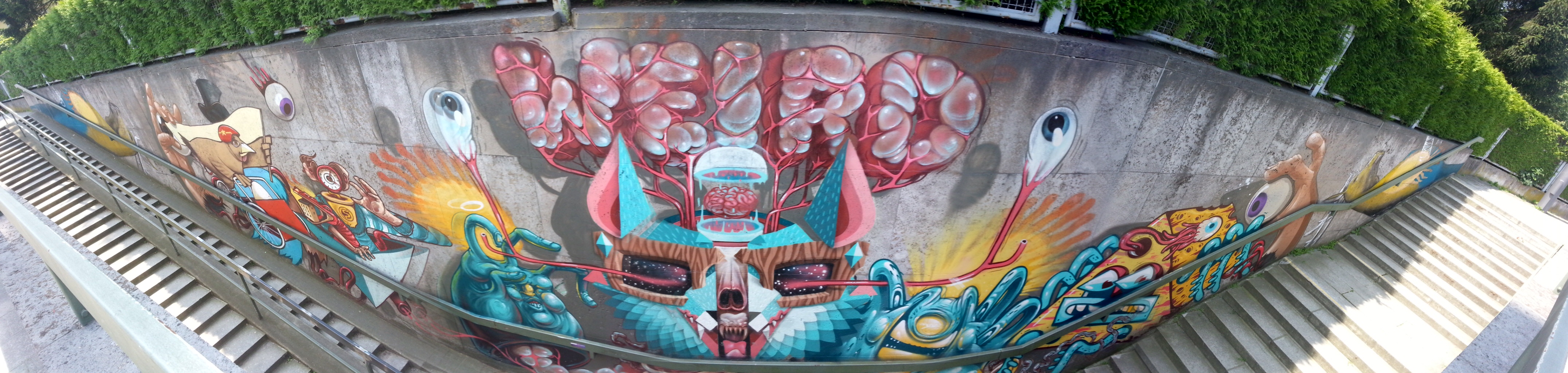
Anatomy terminology hidden in 3D block letters,

Picture1

Hominin canines in Kazakhstani baursaki cakes,

Picture1

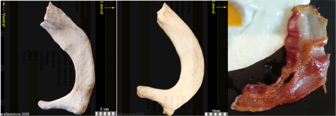
The Ardipithecus ramidus ilium in Almaty,
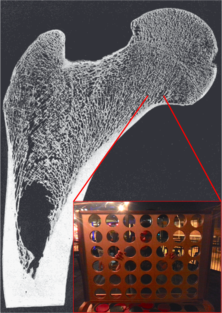
screen-shot-2016-11-26-at-5-36-05-pm

Homo naledi juvenile femur head in nutmeg,

bangkok-erectus

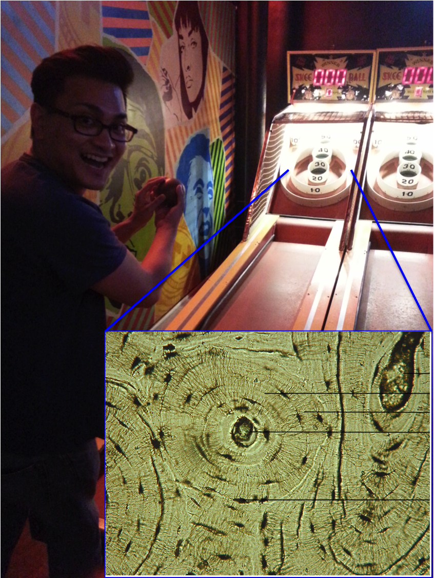
And a Homo erectus cranium on a Bangkok sidewalk. As I’m teaching a fossil-focused seminar next semester, OA will probably become increasingly about fossils, and I’ll probably get my students involved in the fun as well.

New discoveries and enduring questions

The most-read post on the blog this year was about the recovery of the oldest human Nuclear DNA, from the 450,000 year old Sima de los Huesos fossils. My 2013 prediction that nuclear DNA would conflict with mtDNA by showing these hominins to be closer to Neandertals than Denisovans was shown to be correct.

giphy

These results are significant in part because they demonstrate one way that new insights can be gained from fossils that have been known for years. But more intriguingly, the ability of researchers to extract DNA from exceedingly old fossils suggests that this is only the tip of the iceberg.

The other major discoveries I covered this year were the capuchin monkeys who made stone tools and the possibility that living humans and extinct Neandertals share a common pattern of brain development.

Pride & Predator

An unrelated image from 2016 that makes me laugh.

The comparison between monkey-made and anthropogenic stone tools drives home the now dated fact that humans aren’t the only rock-modifiers. But the significance for the evolution of human tool use is less clear cut – what are the parallels (if any) in the motivation and modification of rocks between hominins and capuchins, who haven’t shared a common ancestor for tens of millions of years? I’m sure we’ll hear more on that in the coming years.

In the case of whether Neandertal brain development is like that of humans, I pointed out that new study’s results differ from previous research probably because of differences samples and methods. The only way to reconcile this issue is for the two teams of researchers, one based in Zurich and the other in Leipzig, to come together or for a third party to try their hand at the analysis. Maybe we’ll see this in 2017, maybe not.

There were other cool things in 2016 that I just didn’t get around to writing about, such as the publication of new Laetoli footprints with accompanying free 3D scans, new papers on Homo naledi that are in press in the Journal of Human Evolution, and new analysis of old Lucy (Australopithecus afarensis) fossils suggesting that she spent a lifetime climbing trees but may have sucked at it. But here’s hoping that 2017 tops 2016, on the blog, in the fossil record, and basically on Earth in general.

Osteology Everywhere: Skeletal Spice

The American winter holiday season is steeped in special spices, such as nutmeg, cloves, cinnamon, and whatever the hell pumpkin spice is. I guess as part of the never-ending War on Christmas, each year this sensory and commercial immersion begins earlier and earlier. Since these have become old news, I’d pretty much forgotten about the seasonal spicecapade until just the other day. In prep for minor holiday gluttony, I was grinding fresh nutmeg when I made a startling discovery. Nutmeg is not just the fragrant fruit of the Myristica fragrans tree. No, there’s something far more sinister in this holiday staple.

download

Merely nutmeg?

The ground section looks superficially like an unfused epiphyseal surface, whereas the rounded outer surface is more spherical. It turns out, in the most nefarious of all holiday conspiracies since the War on Christmas, nutmeg halves are nothing more than unfused femur heads! Compare with the epiphyseal surface of this Homo naledi femur head:

screen-shot-2016-11-26-at-5-36-05-pm

Nutmeg (left) and H. naledi specimen UW 101-1098 (right).

This immature H. naledi specimen was recently published (Marchi et al., in press), and the associated 3D surface scan has been available for free download on Morphosource.org for a while now. It fits onto a proximal femur fragment, UW 101-1000, also free to download from Morphosource.

screen-shot-2016-11-26-at-5-48-42-pm

Modified Fig. 11 from Marchi et al. It’s weird that only H. naledi bones were found in the Dinaledi chamber, but even weirder is the underreported presence of nutmeg.

Like most  bones in the skeleton, the femur is comprised of many separate pieces that appear and fuse together at different, fairly predictable ages. The shaft of the femur appears and turns to bone before birth, and the femur head, which forms the ball in the hip joint, usually appears within the first year of life and fuses to the femur neck in adolescence (Scheuer and Black, 2000). So we know this H. naledi individual was somewhere between 1–15ish years by human standards, probably in the latter half of this large range.

So there you have it. Osteology is everywhere – the holidays are practically a pit of bones if you keep your eyes open.

ResearchBlogging.orgREFERENCES

Marchi D, Walker CS, Wei P, Holliday TW, Churchill SE, Berger LR, & DeSilva JM (2016). The thigh and leg of Homo naledi. Journal of Human Evolution PMID: 27855981.

Scheuer L and Black S. 2000. Developmental Juvenile Osteology. New York: Elsevier Academic Press.

Osteology Everywhere: Vertebral Incidens

Try as I might, I can never escape osteology. Never. Just the other day, I was walking through my school’s expansive, boneless atrium, when these haphazardly scattered letters stopped me in my tracks:

2016-02-09 16.26.31

DЯSTUDENSN

Amidst this alphabet soup, there it was, calling out to me. Whispering. Longing….

Untitled

Ah, the dens. What is the “dens” you ask? It is a special little projection on a special little bone, the second cervical vertebra (C2). Why is it special? Well, most vertebrae look pretty similar to one another, with a body in the front being held in awkward embrace by a bony neural arch in the back.


But not the first two vertebrae, C1 and C2. No, these rebels are spinal celebrities. C1, whose rock name is “Atlas” (presumably in honor of its favorite episode of Wishbone) cradles the skull’s occipital condyles on its concave shoulders. Lacking a true body or centrum, Atlas viewed from the top resembles the gaping maw of a manta ray:

Top: Manta ray. Bottom: Atlas viewed from top, anterior is on the bottom (from Scheuer and Black, 200). A and F refer to the age at which the bony portions appear and fuse, respectively.

Top: Manta ray. Bottom: Atlas viewed from top, anterior is on the bottom (from Scheuer and Black, 2000). A and F refer to the age at which the bony portions appear and fuse, respectively.

Atlas is a jerk and so it sits right on top of C2, whose rock name is Axis (after the second album by the Jimi Hendrix Experience). More gawky and angsty than Atlas, Axis differs from the rest of the vertebrae in having an extension, the dens, which reaches skyward to boop the inside of Atlas’ maw:

Top: Axis viewed from the front. Bottom: Axis getting pwnd by Atlas. Modified from White et al. 2012.

Top: Axis viewed from the front. Bottom: Axis getting pwnd by Atlas. Modified from White et al. (2012).

The most distinctive feature of Axis, aside from its smoldering adolescent rage, is the dens (or odontoid process). If you find a bone fragment that is verily vertebral and has a perpendicular projection, you can bet good tenge you’ve got an Axis. Even a densless fragment can be distinguished from all other vertebrae by its superior articular facets, which are rather flat and face mostly superiorly.

What I thought would be a casual jaunt after class last week turned out to be a horrific reminder of the most amazing vertebrae. This must be  how Scott Williams always feels.

Osteology Everywhere: Bacon or first rib?

I went to a cafe today to eat breakfast and get some work done. Write, write, write. It’s important to be properly nourished to ensure maximal productivity.

The Ron Swanson diet.

The Ron Swanson diet.

But I was aghast to behold the food they placed before me:

More bacon, please.

What on earth is this?

First of all, this is not a sufficient amount of bacon.

ljQEkGT

Secondably, this bacon is a spitting image of a first rib:

First ribs, from left to right: Human, chimpanzee, bacon. First two images from eSkeletons.org.

First ribs from the right side of the body, viewed from the top. From left to right: Human, chimpanzee, bacon. First two images from eSkeletons.org.

At the top of the ribcage, just beneath the clavicle and subclavian artery and vein, the first rib is much shorter and flatter than the rest of the ribs. As Jess Beck at Bone Broke points out, “The first and second rib give something of an awkward ‘slow song at a middle-school dance’ kind of a hug, while the lower ribs provide a more comfortable and self-assured embrace.” I mean, just lookit how sheepishly the bacon dances with the eggs in the first picture, it has ‘middle-school dance’ written all over it.

But the bacon is not totally identical to the human and chimpanzee counterparts. It’s missing their anteromedially sweeping arc, and the distal portion reaching out to the egg is fairly straight. This suggests we’re probably missing much of the original distal end. Posteriorly or dorsally (toward the bottom in the pic), it also appears to be missing much of the lateral portion including the vertebral facet. In this regard, this bacon rib looks a lot like the first rib of Homo naledi:

Full stack of ribs. From left to right: Human, bacon, Homo naledi, Dmanisi Homo erectus, Australopithecus sediba (x2), Australopithecus afarensis specimen "Lucy," Ardipithecus ramidus, and chimpanzee. Images not to scale except Lucy and Ardi.

Full stack of ribs. Left to right: Human, bacon, Homo naledi, Dmanisi Homo erectus, Australopithecus sediba (x2), Australopithecus afarensis specimen “Lucy,” Ardipithecus ramidus, and chimpanzee. Images not to scale except Lucy and Ardi. Image credits given below.

It is hard to make good homologous comparisons among these fossils and bacon, since so many are so incomplete. But it looks like the hominins are relatively longer (front to back, or dorsoventrally) compared to the chimpanzee. That is, oriented along the rib “neck,” the ventral/distal end projects far more medially beyond the proximal vertebral facet in the chimp, while in the hominins the two ends are more flush.  Ardi is really incomplete and so very hard to orient, but it may be more like the chimp (I think it needs to be rotated to the right more, to make the lateral edge more vertical like all the other specimens).

It will be interesting to see what my colleagues working on the Homo naledi thorax have to say about rib shapes and their functional importance, hopefully not too long from now.

Anyway, I really wish I had more bacon.

Fossil rib sources
ResearchBlogging.orgDmanisi Homo erectus: Lordkipanidze D, Jashashvili T, Vekua A, Ponce de León MS, Zollikofer CP, Rightmire GP, Pontzer H, Ferring R, Oms O, Tappen M, Bukhsianidze M, Agusti J, Kahlke R, Kiladze G, Martinez-Navarro B, Mouskhelishvili A, Nioradze M, & Rook L (2007). Postcranial evidence from early Homo from Dmanisi, Georgia. Nature, 449 (7160), 305-10 PMID: 17882214

Australopithecus sediba: Schmid P, Churchill SE, Nalla S, Weissen E, Carlson KJ, de Ruiter DJ, & Berger LR (2013). Mosaic morphology in the thorax of Australopithecus sediba. Science, 340 (6129) PMID: 23580537

Homo naledi: Morphosource.

Australopithecus afarensis and Ardipithecus ramidus: White TD, Asfaw B, Beyene Y, Haile-Selassie Y, Lovejoy CO, Suwa G, & WoldeGabriel G (2009). Ardipithecus ramidus and the paleobiology of early hominids. Science, 326 (5949), 75-86 PMID: 19810190

Osteology Everywhere: Why we number our premolars 3-4

Portishead* came on the radio the other day, making iTunes display the cover of their album, Third. My inner osteologist rejoiced to see it prominently features a tooth!

Third album cover by Porthishead (2008). Image from Wikipedia

Well not a picture, but rather the name, of a tooth. In each quadrant of your mouth (most likely) are two premolars, commonly referred to as “bicuspids.” In the biz, we usually call these pals,  “P3” and “P4.”

UW 101-1277 mandible, part of the Homo naledi holotype skull. Modified from the Wits media gallery.

UW 101-1277 mandible, part of the Homo naledi holotype skull. Each capital letter stands for the tooth type (incisor, canine, premolar, and molar). Modified from Wits’ image gallery.

You might be wondering why we call them P3 and P4, when there are only two premolars per quadrant — what happened to P1 and P2?  Homology to the rescue!

The ancestral mammalian condition was to have four premolars (and a 3rd incisor) in each side of the jaw. This is a “dental formula” of 3-1-4-3, indicating the numbers of each tooth type from front to back. Over time, different groups of animals have lost some of these teeth. Baleen whales have lost all of them.

P1 and an incisor were lost early in the evolution of Primates. Most Strepsirrhines and New World monkeys retain this primitive”2-1-3-3″ dental formula :

Ring tailed lemur (left) and woolly monkey (right) maxillae, showing the primitive primate dental formula including a P2. For scale, gridlines are 10 mm (left) and 20 mm (right).

Ring tailed lemur (left) and woolly monkey (right) maxillae, showing the primitive primate dental formula including a P2. For scale, gridlines are 10 mm (left) and 20 mm (right). Images from this boss database.

The last common ancestor of catarrhines (living humans, apes and Old World monkeys) lost the P2, and so we have only two premolars left in each side of the jaw. These are homologous with the third and fourth premolars of the earliest mammals. And that’s why we call them P3-4.

*The song was “The Rip.” It’s a very good song with an insanely creepy and trippy video:

Osteology Everywhere: Vertebeer Fest

This past weekend was witness to the Summer Beer Festival, the annual showcase of Michigan’s brewing splendor. Dozens of breweries brought out batches of beer, from classics we know and love, to inspired innovations meriting a MacArthur Fellowship. There was an embeerrassment of boozes. Dark Horse Brewing Company, from Marshall, MI, put on quite the show:

Dark  Horse Brewing Co. pumping out the brews and blasting t-shirts into the crowd.

Dark Horse Brewing Co. pumping out the brews and blasting t-shirts into the crowd.

Besides towering over the bacchanal hordes, the Dark Horse beer fort also offered IPAs infused with pretty much anything that might pair well with hops. They even steeped habañero peppers in one, and it was maximally boss.

Beer still my heart.

Beer still my heart.

Having sampled only a small part of rich the smorgasbord on tap, a rest by the river was in order. The Festival was on the banks of the mighty Huron River, an excellent place to sit and sip Arcadia‘s scotch ale, taking in the evening under cloud-peppered, cerulean skies. Such a calm and relaxing setting would surely offer respite for a brain besieged by bones. Right?

Every year for the Festival they replace the river water with beer.

Every year for the Festival they replace the river water with beer.

Wrong! Peering through beer goggles over the shimmer of the river, seeking signs of Bigfoots lurking on the opposite shore, I locked eyes with a large, wooden vertebral body.

No ordinary tree stump

An eyeless frown marks the ventral surface of this centrum.

The human spine is composed of anywhere from 31-34 vertebrae (not counting the coccyx or tail bone). The body or “centrum” is the large, blocky portion of the bone, which is separated from other such bodies by intervertebral discs; it is literally a pile of bodies, stacked one on top of the other. And the intervertebral discs are remnants of the notochord, the embryonic structure that unites you and me and all other humans with all other animals known as chordates. Anyway, kiss my grits if this old tree stump across the mighty Huron River here doesn’t look like a lower thoracic or upper lumbar vertebral body, the metaphoric shark fin of a giant trunkless human waiting to pounce from the placid waters.

a) Our mystery vertebra. b) a lumbar vertebra from White et al. (2012). c) views of the right and front side of the Australopithecus africanus fossil StW H41, from Sanders (1998, Fig. 1).

a) Our mystery river vertebra. b) a lumbar vertebra from White et al. (2012). c) views of the right and front side of the Australopithecus africanus fossil StW H8/H41, modified from Fig. 1 of Sanders (1998).

Thinking on it, our mystery river vertebra doesn’t just look like any old human centrum, it is a ringer for the second lumbar vertebra of StW H8/H41, a series of the 11th thoracic to 4th lumbar vertebrae of Australopithecus africanus from Sterkfontein (see the red arrow in c, above). Sanders (1998) notes that this short segment of an early hominin spine shows clear adaptation to walking upright like we humans do today, although the size of the vertebral bodies is both absolutely and relatively small compared to ours, just as is seen in other Australopithecus fossils.

And what better way to celebrate this monumental discovery than returning to the Beer Festival – hooray beer!

Osteology Everywhere: Barcade bone biology

I’ve fled the Central Asian steppe to visit my childhood home, Kansas City, Missouri.

The tortuous path from the center of Eursia to the center of the US, a mere 8500 miles since there are no direct flights. Map made by Wolfram Alpha.

The tortuous path from the center of Eursia to the center of the US, a mere 8500 miles since there are no direct flights. Map made by Wolfram Alpha.

It would be a lie to say I don’t miss life in this Midwest metropolis. Kansas City is sprawling, with diverse cultures, foods and festivities in far-flung neighborhoods. It’s always a trip to revisit the people and places of my formative years.

Of course, there are differences between now and when I was growing up. A whole new world of experiences became available to me here once I was old enough to drink (legally; this is long ago now). The bar scene itself has evolved over the past decade or so, arguably culminating in Up-Down, a grown-up video game arcade that will confusingly make you both happy and sad to have become an adult.

Be still my heart. Image credit.

I’ve never seen anything like this before. But even in this novel environment, I still couldn’t help but notice Osteology Everywhere. What appears at first glance to be an oversized Connect Four contraption . . .

Go for the bottom, go for the top.

Go for the bottom, go for the top.

. . . is in fact a closeup of trabecular bone (with my friend creepily peering through):

Section through a human proximal femur (hip joint). Note the trabecular or "spongy" bone filling the top, in comparison with the thick and dense cortical bone of the shaft in the bottom left. Image credit.

Vertical section through a human proximal femur (hip joint). Note the trabecular or “spongy” bone filling the top, in comparison with the thick and dense cortical bone of the shaft in the bottom left. Image credit.

And here, we’re not playing Skee Ball . . .

20150618_003612. . . we’re hurling wooden balls into Haversian canals and lacunae of osteons. For Science.

Cross section through cortical bone, magnified to highlight an osteon. The big hole in the center is the Haversian canal, and the smaller satellite holes are lacunae housing osteocytes.

Cross section through cortical bone, magnified to highlight an osteon. The big hole in the center is the Haversian canal, and the smaller satellite holes are lacunae housing osteocytes. Image credit.

So if you’re in the KC area, I highly recommend you check out Up-Down, where you can review osteology while also playing games and sipping a refreshing beer. Who knew learning could be so fun?

The stream-severed spine

I recently returned from Mangystau, a geologically captivating former seabed in West Kazakhstan. Places like this, or the Tien Shan mountains in the South and Altai mountains in the East, always make me wonder why anyone would decide to build a capital city in the wastes of Aqmola. Astana sprouts up from a sterile steppe, sparingly sprinkled with streams and lakes. Out west, though, are breathtaking landscapes and landforms, such as the giant rocky spheres of Torysh:

Traversing the

Traversing the “Valley of Balls.” It is not yet known what caused these rock formations.

Sherqala (“Lion City”), a rocky uplift that centuries ago hosted a defensive acropolis:

DSC01089

A flooded salt flat vertiginously reflecting an alternate reality:

DSC01226
A pile of earth that’s really an octopus waiting in ambush:

DSC01315

The perfect place to set up camp.

DSC01400

As I’d pointed out the first time I came out here last year, this rocky terrain is littered with lifeless remnants of the animals that used to call this place home. So many bones reflecting such biodiversity, just lying on the surface. This year, though, I found a subsurface skeleton, teaching an important lesson in taphonomy. Taphonomy (“burial law” from Ancient Greek) is the study of what happens to an animal’s remains from the moment it dies to when it is discovered eons later. This field examines geological and ecological processes that determine whether fossils are found intact or smashed to smithereens.

Walking down into a small gully by our campsite, I noticed some giant lumbar vertebrae eroding out of one side:

Waist-deep in mud. One vertebra is clearly visible, and to its right, beneath a rock, are the spinous processes of two more vertebrae.

Waist-deep in mud. One vertebra is clearly visible, and to its right, beneath a rock, are the spinous processes of two more vertebrae. Notice the differently colored stripes of soil – these are different layers (“strata”), reflecting different periods that soil was laid down on the earth.

I was elated to espy this spinous surprise, but I wasn’t expecting to see what was on the opposite side of the gully:

Died doing a misguided impression of an ostrich.

Died doing a misguided impression of an ostrich. On the left you can see the back of the skull and the first cervical vertebra, then the spine submerges and reemerges to the right.

Sure enough, this once complete carcass was drawn and quartered, pulled apart by the liberal application of time and life-saving water.

Digging out the skull on the west bank, right across from the lumber spine on the east face. The different soil layers (

Digging out the skull on the west bank, right across from the lumber spine on the east face (circled).

Getting our hands a little dirty, we found the face of a camel. It is hard to say how long ago it lived, how long it took to get buried by a few inches of dirt, but I would guess at most only a few decades (but I’m not a geologist, so who knows). It’s also unclear how this animal was bifurcated: Did the camel die and get covered over with soil, and then later a newly forming stream carried away the soil harboring its torso? Or did the carcass lie on the ground unburied for a while, its torso slowly picked apart or trampled, and then the stream formed? I would guess the first scenario is more likely, since the bones seem to run through several strata. But again I’m not an expert in taphonomy so I could be wrong.

DSC01023

People often wonder why the fossil record isn’t more complete, and why we get so excited about the discovery of even partially complete skeletons. This camel demonstrates one of myriad taphonomic processes, one of the many ways that earth, water and time conspire tear the past asunder.

Osteology everywhere: Graffiti

Astana, the wedding-cake capital of Kazakhstan, is notably bereft of graffiti and street art, at least in my somewhat limited exposure to the city. The larval metropolis is all about commercial appearance, so I’d guess that aspiring street artists likely face much more than the Marge Simpson treatment for turning around to brag about their work.

Dire consequences await those who graffito tag public property.

Dire consequences await those who graffito tag public property.

Once, I did see a pretty badass street mural,

But it was in München.

but it was in München, a mere 2,620 miles from Astana.

No, there is not much in the way of secretly donated street art here in Astana, and there’s generally little hope to see graffiti-grafted Osteology Everywhere. But this weekend, I noticed these four magical letters, quickly quietly scrawled on the side of my apartment building:

2015-01-17 14.34.37

DAKA.

Two disconcerting thoughts immediately come to mind reading this. First, why the hell is “DAKA” written in Latin instead of Cyrillic script characteristic of the FSU? Second, what does “DAKA” mean out here? Nothing in Russian so far as I know, but Google Translate claims it could mean “Dakar” in Kazakh, which if true raises even more questions.

No, the safest assumption is that this tagger, my streetwise and marker-wielding dopplegänger, was referring to the ~1 million year old Homo erectus partial skull from Ethiopia, dubbed “Daka” after the Dakanihylo site of its discovery.

The Daka calvaria (Figure 2. of Asfaw et al., 2002). Counterclockwise from the top left: view from the back, view from the top (front is to the left), view from the left, a mosquito net, view from the bottom (front is at the top), viewed from the front.

BOU-VP-2/66, the Daka calvaria* (Figure 2. of Asfaw et al., 2002). Counterclockwise from the top left: view from the back, view from the top (front is to the left), view from the left, a mosquito net, view from the bottom (front is at the top), and view from the front. *Calvaria is the fancy word for ‘bony skull without a face.’

Daka isn’t the first hominin fossil to be embraced outside of anthropology. A few years ago I noticed the 4.4 million year old Ardipithecus ramidus skeleton strutting across the label of a Dogfish Head beer bottle:

2011-11-12_14-41-32_247

GOODGRIEF, this was almost 5 years ago.

In downtown Tbilisi, Georgia I recently spotted a Dmanisi-based duo whose tech savvy belies the fact they’re based on 1.8 million year old fossils:

20141013_141725

(Let’s not forget this one, from before they got smartphones)

We’ll have to do some serious fossil-finding here in Kazakhstan before they’ll let anyone put up something this awesome on the side of anything here in Astana. (Or wait…)

Osteology everywhere: Pollicem verte(b)r(a)e [Latin puns are hard]

I just got back from the meetings of the European Society for the Study of Human Evolution in Florence. As you can guess, bones and genes and anatomy and apes and biomechanics and energetics and everything were on everyone’s minds. Even in the midst of an unseasonal surprise typhoon of lunch time ice:

Ambush of hail.

Aw hail no.

Along the way, I passed a gift shop window and this book cover immediately caught my eye:helert

No, it’s not an ancient Roman gladiator’s helmet. It’s clearly a lumbar vertebra, probably of some quadruped. We’re looking down onto the top (or front of it) from the cranial view. The body or centrum is the rounded part toward the bottom of the picture, the short transverse processes jutting off to the sides. The spinous process, pointing toward the top, is even thick and blunt distally as is characteristic of lumbar verts. Here’s a comparison:

Middle lumbar vertebrae, from the cranial view (modified from Figs. 3-4 of Moyà-Solà et al., 2004). 0=modern baboon, A=Proconsul nyanzae (KNM-MW 13142-J)(B) P. catalaunicus (IPS-21350.59). (C) Cast of Morotopithecus bishopi (UPM 67.28) from Moroto (Uganda). (D) D. laietanus (IPS-18000) from Can Llobateres (Spain). (E) Pongo pygmaeus

Middle lumbar vertebrae of various Miocene apes (A-D) in cranial view (modified from Figs. 3-4 of Moyà-Solà et al., 2004). 0=modern baboon, A=Proconsul nyanzae (KNM-MW 13142-J), B=Pierolapithecus catalaunicus (IPS-21350.59), C=Morotopithecus bishopi (UPM 67.28), D=Hispanopithecus laietanus (IPS-18000), and E= modern orangutan.

Modern apes use an upright posture more frequently than living monkeys, who are quadrupedal. An anatomical correlate of these postures is the position of the transverse processes. Compare the baboon (0 in the figure above) with the orangutan (E). In the monkey the transverse processes come off the sides of the centrum (below the horizontal line), while in the orangutan the processes come off the pedicle further back. In your lumbars the transverse processes arise a little bit more toward the back than in the orangutan.

This is a pretty characteristic pattern, meaning that we can reconstruct the habitual posture of an animal based on a single bone – even just part of a single bone as in the case of Hispanopithecus (D, above). Proconsul nyanzae (A), dating to around 19 million years ago and therefore one of the earliest apes, has a monkey-like lumbar vert; the rest of its skeleton is monkey-like and so we think many of the earliest apes moved around like modern monkeys. In contrast, Morotopithecus bishopi (C), at 20.6 million years ago, is also one of the earliest apes but has a more modern-ape-like lumbar. And so with Pierolapithecus and Hispanopithecus.

The vertebra gracing the cover of our gift shop book is clearly more monkey-like, presumably from a simian who long ago walked on all fours across the blood-soaked floors of a cacophonous Colosseum.