Osteology Everywhere: Ilium Nublar

Jurassic Park is objectively the greatest film ever made, so I don’t need to explain why I recently watched it for the bajillionth time. Despite having seen this empirically excellent movie countless times, I finally noticed something I’d never seen before.

Hold on to your butts. What's that on the screen in front of Ray Arnold?

Hold on to your butts – what’s that on the screen in front of John Arnold? (image credit)

The film takes place on the fictitious island “Isla Nublar,” a map of which features prominently in the computer control room when s**t starts to go down. Here’s a clearer screenshot of one of Dennis Nedry‘s monitors:

Isla Nublar from the JP control room. Quiet, all of you! They’re approaching the tyrannosaur paddock…. (image credit)

It dawned on me that the inspiration for this island is none other than MLD 7, a juvenile Australopithecus africanus ilium from the Makapansgat site in South Africa:

Figure 1 from Dart, 1958. Left side is MLD 7 and right is MLD 25. Top row is the lateral view (from the side) and bottom row is the medial view (from the inside).

Figure 1 from Dart, 1958. Left side is MLD 7 and right is MLD 25. Top row is the lateral view (from the side) and bottom row is the medial view (from the inside). These two hip bones are from the left side of the body (see the pelvis figure in this post). Note the prominent anterior inferior iliac spine on MLD 7, a quintessential feature of bipeds.

Isla Nublar is basically MLD 7 viewed at an angle so that appears relatively narrower from side to side:

MLD at a slightly oblique view (or stretched top to bottom) magically transforms into Isla Nublar.

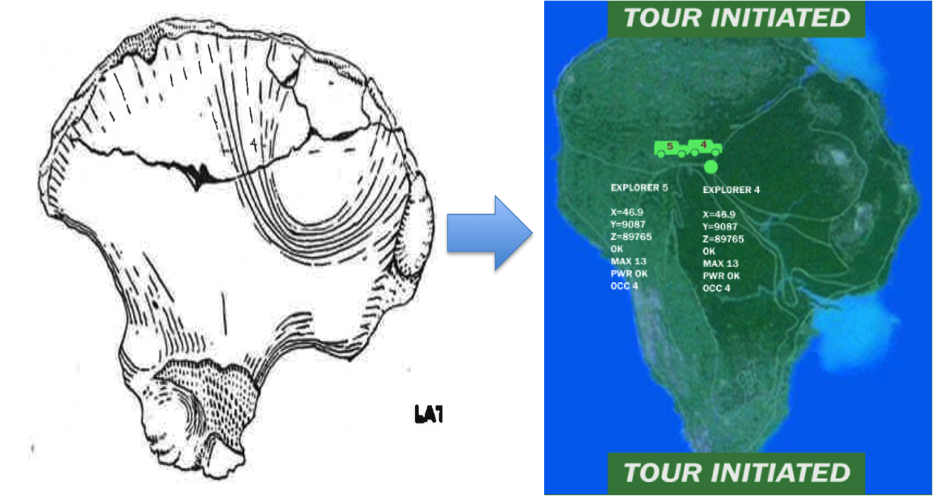
MLD 7 at a slightly oblique view (or stretched top to bottom) magically transforms into Isla Nublar.

It’s rather remarkable that some of the most complete pelvic remains we have for australopithecines are two juveniles of similar developmental ages and sizes from the same site. In both, the iliac crest is not fused, and joints of the acetabulum (hip socket) hadn’t fused together yet. The immaturity of these two fossils matches what is seen prior to puberty in humans and chimpanzees. Berge (1998) also noted that MLD 7, serving as an archetype for juvenile Australopithecus, is similar in shape to juvenile humans, whereas adult Australopithecus (represented by Sts 14 and AL 288) are much flatter and wider side to side. Berge took this pattern of ontogenetic variation to match an ape-like pattern of ilium shape growth. This suggests a role of heterochrony in the evolution of human pelvic shape, or as Berge (1998: 451) put it, “Parallel change in pelvic shape between human ontogeny and hominid phylogeny.” In layman’s terms, ‘similar changes in both pelvic growth and pelvis evolution.’

Gona … Gona … not Gona work here anymore more

The Gona pelvic remains (A-D), and the reconstructed complete pelvis (E-J), Fig. 2 in Simpson et al., 2008.

A few years ago, Scott Simpson and colleagues published some of the most complete fossil human hips (right). The fossils are from the Busidima geological formation in the Gona region of Ethiopia, dated to between 0.9-1.4 million years ago. (Back when I wasn’t the only author of this blog, my friend and colleague Caroline VanSickle wrote about it here)

Researchers attributed the pelvis to Homo erectus on the basis of its late geological age and a number of derived (Homo-like) features. In addition, the pelvis’s very small size indicated it probably belonged to a female. One implication of this fossil was that male and female H. erectus differed drastically in body size.

Christopher Ruff (2010) took issue with how small this specimen was, noting that its overall size is more similar to the small-bodied Australopithecus species. Using the size of the hip joint as a proxy for body mass, Ruff argued Gona’s small size would imply a profound amount of sexual dimorphism in H. erectus: much higher than if Gona is excluded from this species, and higher than in modern humans or other fossil humans. Ruff thus proposed an alternative hypothesis to marked sexual dimorphism, that the Gona pelvis may have belonged to an australopithecine.

Fig. 3 From Ruff's (2010) reply. Australopiths (and Orrorin) are squares and Homo are circles. Busidima's estimated femur head diameter is represented by the star and bar.

Fig. 3 From Ruff’s (2010) reply. Australopiths (and Orrorin) are squares and Homo are circles. Gona’s estimated femur head diameter is represented by the star and bar.

Now, Simpson & team replied to Ruff’s comments, providing a laundry list of reasons why this pelvis is H. erectus and not Australopithecus. They cite many anatomical features of the pelvis shared with Gona and Homo fossils, but not australopithecines. They also note that there are many other bones reflective of body size, that seem to suggest a substantial amount of size variation in Homo fossils, even those from a single site such as Dmanisi (Lordkipanadze et al., 2007).

Interestingly, neither of these parties compared the implied size variation with that of living apes. So I’ll do it! Now, I do not have any acetabulum data, but a friend lent me some femur head measurements for living great apes a few years ago. Gona is a pelvis and not a femur, but there are more fossil femora than hips. Because there’s a very high correlation between femur head and acetabulum size, Ruff estimated Gona’s femur head diameter to be 32.6 mm (95% confidence interval: 30.1-35.2; Simpson et al. initially estimated 35.1 mm based on a different dataset and method). To quantify size variation, we can compare ratios of larger femur heads divided by smaller ones. Now, this ratio quantifies inter-individual variation, but it will underestimate sexual dimorphism since I’m likely sampling some same-sex pairs that aren’t so different in size. But this is just a quick and dirty look. So, here’s a box plot of these ratios for Homo fossils, larger specimens divided by Gona’s estimated femur head size in different time periods:

Ratio of a fossil Homo femur head diameter (HD) divided by Busidima's HD. E Homo = early Pleistocene, Contemporaneous = WT 15000 and OH 28, MP = Middle Pleistocene Homo. White boxes are based on Ruff's Busidima HD estimate, green boxes are based on Simpson et al.'s estimate.

Ratios of fossil Homo femur head diameter (HD) divided by Busidima’s (Gona’s) HD. E Homo = early Pleistocene, Contemporaneous = WT 15000 and OH 28, MP = Middle Pleistocene Homo. White boxes are based on Ruff’s Gona HD estimate, green boxes are based on Simpson et al.’s larger estimate. Boxes include 50% quartiles and the thick lines within are sample medians.

Clearly, Gona is much smaller than most other fossil Homo hips, since ratios are never smaller than 1.14. Average body size increases over time in the Homo lineage, reflected in increasing ratios from left to right on the plot. Early Pleistocene Homo fossils are fairly small, including Dmanisi, hence the lower ratios than later time periods. Middle Pleistocene Homo (MP), represented by the most fossils, shows a large range of variation, but even the smallest is still 1.17 times larger than the largest estimate of Gona’s femur head size. To put this into context, here are those green ratios (assuming a larger size for Gona) compared with large/small ratios from resampled pairs of living apes and humans:

*

The fossil ratios of larger/smaller HD from above, compared with resampled ratios from unsexed living apes and humans. Boxes include the 50% quartiles, and the thick lines within are sample medians. **(05/03/14: This plot has been modified from the original version post, which only included the fossil ratios based on the smaller Gona estimate)

What we see for the extant apes and humans makes sense: humans and chimpanzees show smaller differences on average, whereas average differences between gorillas and orangutans are larger. This accords with patterns of sexual dimorphism in these species. **What this larger box plot shows is that if we accept Ruff’s smaller average estimate of Gona’s femur head size (white boxes), it is relatively rare to sample two living specimens so different in size as seen between Gona and other fossils. If we use Simpson et al.’s larger Gona size estimate, variation is still elevated above most living ape ratios. Only when Gona is compared with the generally-smaller, earlier Pleistocene fossils, does the estimated range of variation show decent overlap with living species. Even then, the overlap is still above the median values.

These results based on living species agree with Ruff’s concern, that including Gona in Homo erectus results in an unusually large range of variation in this species. Such a large size range isn’t necessarily impossible, but it would be surprising to see more variation than is common in gorillas and orangutans, where sexual size dimorphism is tremendous. Ruff suggested that the australopith-sized Gona pelvis may in fact be an australopith. This was initially deemed unlikely, in part because the fossil is well-dated to relatively late, 0.9-1.4 million years ago. However, Dominguez-Rodgrigo and colleauges (2013) recently reported a 1.34 mya Australopithecus boisei skeleton from Olduvai Gorge, so it is possible that australopiths persisted longer than we’ve got fossil evidence for, and Gona is one of the latest holdouts.

So many possible explanations. More clarity may come with further study of the fossils at hand, but chances are we won’t be able to eliminate any of these possibilities until we get more fossils. (also, the post title wasn’t a jab at the fossils or researchers, but rather a reference to the movie Office Space)

References

Dominguez-Rodrigo et al. 2013. First partial skeleton of a 1.33-million-year-old Paranthropus boisei from Bed II, Olduvai Gorge, Tanzania. PLoS One 8: e80347.

Ruff C. 2010. Body size and body shape in early hominins – implications of the Gona pelvis. Journal of Human Evolution 58: 166-178.

Simpson S et al. 2008. A female Homo erectus pelvis from Gona, Ethiopia. Science 322: 1089-1092.

Simpson S et al. In press. The female Homo pelvis from Gona: Response to Ruff (2010). Journal of Human Evolution. http://dx.doi.org/10.1016/j.jhevol.2013.12.004