In the Summer of 2019 I worked with some great Vassar undergrads to make virtual endocasts and generate new brain size estimates for the Neandertals from the site of Krapina, which we then published in 2021 (discussed in this blog post). The virtual approach to endocast reconstruction uses 3D landmark-based geometric morphometrics methods, and so in the spirit of open science we also published all the landmark data used for the study (as well as a bunch of other fossil human brain size estimates) in the Zenodo repository (here).
Neandertal fossil specimens Krapina 3 (purple/green) and Krapina 6 (yellow/red) with preserved landmarks and virtually reconstructed endocasts.
Something major and global happened around that time — who can even remember what? — and so I never got around to posting R code to accompany the study. So, I’ve finally gotten around to adding some very basic code to the Zenodo entry (better late than never). The code simply reads in the landmarks, estimates missing data for fossils, and does some very basic shape analysis and visualization. It’s doesn’t get into all the nuts and bolts of our study, but it should be enough to help folks check our data or get started with shape analysis in R.
R code includes ways to visualize the landmark data. Left: Principal components analysis graph of endocast shape for humans (red) and Neandertals (blue). Right: Triangle meshes of the average human and Neandertal endocast shapes, viewed from the right, bottom, and back.
Original article Cofran Z, Boone M, Petticord M. 2021. Virtually estimated endocranial volumes of the Krapina Neandertals. American Journal of Physical Anthropology 174: 117–128. (link)
In the latest paper out of the lab (here), my students and I reconstructed the brain endocasts of the Krapina Neandertals. The Krapina rock shelter in Croatia is a remarkable site. Dating to around 130,000 years ago (if not older), the Krapina fossils are early members of the Neandertal lineage. In addition, the fossils represent dozens of Neandertals, from infants to adults. Part of what drew me to the site were the juvenile skulls, since they can tell us about growth and development in these early humans. But, the fossils are quite fragmentary, and needed to be reconstructed to estimate important characteristics like brain size.
Figure 1 from our paper, showing the five Krapina crania (A & B are the same individual) with the endocranial surface highlighted.
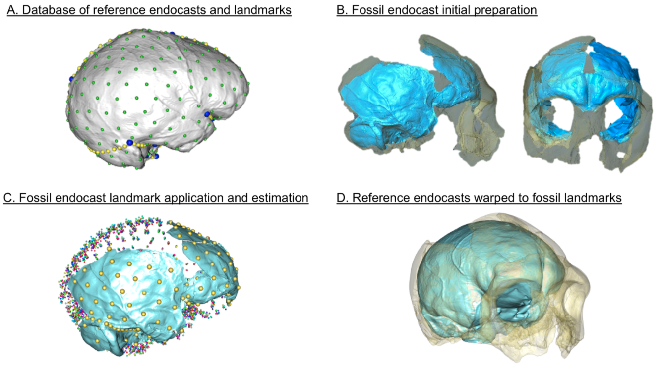
Vassar College has a great program called URSI, where students team up with faculty to get hand on experience conducting research over the summer. So, two summers ago my students and I worked on virtually putting these Humpties Dumpty back together again. Using 3D surface scans of the original fossils and CT scans of modern humans, we used virtual methods to digitally reconstruct the endocasts, which are a good proxy for brain size and shape. Here’s the basic workflow:
Figure 2 from the paper, depicting the workflow for virtually reconstructing fossil endocasts, represented by the famous Krapina 3 or “C” cranium.
The human endocasts were produced from recent humans from the Terry Anatomical Collection, generously made available here by Dr. Lynn Copes. We have posted the 3D landmark data for the humans, the preserved landmarks from the Neandertals, and a big list of estimated brain sizes for Neandertals, in the open access repository Zenodo (here). So, hopefully anyone can repeat our results, or use these data in their own research.
With virtual methods, we could generate multiple reconstructions of each Neandertal fairly easily, giving an idea of how certain or uncertain our brain size estimates were. In the end, we showed that i) the Krapina juveniles, who were probably around 6-7 years old, had brain sizes within the adult range (it’s same with modern humans); ii) average brain size at Krapina was a little lower than previously estimated; and iii) although later Neandertals from other sites had larger brains on average, the difference is not necessarily greater than could be expected by chance.
I’ve participated in Vassar’s URSI program for the past few years and it has been a lot of fun. Last (virtual) summer, my students and I compared hip growth in humans and Australopithecusafricanus, and this coming summer we will examine the brains of the greatest animals of all time — gibbons!