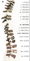
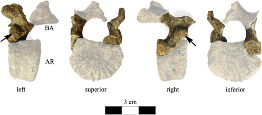
Variation: a blessing and a curse

Trying to start on finishing my dissertation, I’m thinking about the issue dental development and how it relates to skeletal growth. Specifically I’m trying to decide whether I want to analyze my human and Australopithecus robustus samples based on estimates of “dental age,” or if I want to be a bit more cavalier and divide the sample into rougher age categories.

To avoid copyright issues, here’s a crappy picture I drew a few years ago, of the youngest A. robustus jaws. The youngest, “SK 438” is erupting its last baby tooth (bottom right), while the others have their full set of baby teeth, and none of them has its first adult tooth yet. I don’t think I can estimate ages accurately enough to capture the true chronological difference between SK 438 and the rest. Would I be better off just dividing the group into “younger” (SK 438) and “older” (the rest) infants, or even lumping them all together as simply “infants”?

On the one hand, I could assign individuals a chronological age based on a modern referent of known age, at similar stages of dental development. This could allow me to get more fine-scale glimpses into patterns of growth in my samples, but that’s assuming I’ve accurately estimated their ages. Individuals vary in the ages and sizes at which their teeth erupt; a person’s first molar, for example, may erupt at anywhere from 4-8 years of age. How can I estimate an individual’s age in light of such variation? And what if I’m as poor a judge of ages as Dennis Duffy?! Conceivably I could program my analysis to account for error estimation (which in itself could be educational and interesting, but is it worth the trouble?), but this would also add a further source of uncertainty. And it’s like Dwight Schrute said (Michael Scott said), “K-I-S-S: keep it simple, stupid. Great advice, hurts my feelings every time.”

On the other hand, I could divide my sample into coarse age categories – say, putting specimens who’ve attained a given level of dental development in the same group, such as ‘infant, child, juvenile, adolescent, and young adult.’ This method loses the temporal resolution of the first method, but also avoids the possible errors of assigning strict ages I’m pretty sure I would not infer accurately. But, tooth development does not show a clean 1-to-1 relationship with other systems in the body, such as hormonal axes or the bony skeleton. It’s uncertain how accurately kids can be put in any of the above categories (based on general life history variables; Bogin 1999) based on dental development.

Choices, choices.

Variation is a problem for biologists. The theory of evolution was conceived as a way to explain the conundrum of why there is such remarkable variation in the forms of life that Earth is lucky to have harbored. The problem of within-species variation in the relative timing of skeletal and dental development isn’t just a bug-bear for paleoanthropologists. It’s important to medical doctors and pathologists investigating genetically-based developmental disorders, and to epidemiologists looking at aspects of population health, such as the prevalence of growth stunting. It’s also important for forensics specialists who need to use biological clues about the age and identity of crime victims and defendants. I mean, how else would we know whether Jon Voight bit both Kramer and this pencil?

The silver lining, I suppose, on this storm-cloud of biological of variation is that without variation there cannot be evolution. And stasis is boring. If nothing changed since the Cambrian, none of us would be here today. We’d probably be some gross stupid monstrous thing, like this Hallucigenia to the right. It’s the quirks and weird variants that arise randomly, that make evolution possible. If individuals all developed exactly the same, then all organisms through all time would be the exact same, and probably all would have gone extinct as they succumbed to some sinister fate, no new variants would have arisen that may have been able to survive the devastation.

ResearchBlogging.org
So variation is a blessing and a curse. Individual and population variation make it difficult to state norms such as what is “average” or “healthy,” and nothing to be concerned about. Variation is also the magic ingredient of adaptation, without which Life could not survive the randomness inherent in any environment.

Things I cited
Bogin, B. (1999). Evolutionary perspective on human growth Annual Review of Anthropology, 28 (1), 109-153 DOI: 10.1146/annurev.anthro.28.1.109

Also 30 Rock, The Office and Seinfeld. Well done, NBC.

Pictures worth thousands of words and dollars

ResearchBlogging.orgLooking into subdural empyema, which is a meningeal infection you don’t want, I stumbled upon a study from the roaring 1970s – the glorious Nixon-Ford-Carter years – using computerized axial tomography (hence, CAT scan) to visualize lesions within the skull (Claveria et al. 1976). Nowadays people refer to various similar scanning techniques simply as “CT” (for computed tomography, though this is not exactly the same as magnetic resonance imaging, MRI).

It’s pretty amazing how medical imaging has advanced in the 35 years since this study. For example, to the right is a CAT scan from Claveria et al. (1976, Fig. 4). These are transverse images (“slices”) through the brain case, the top of the images corresponding to the front of the face. You can discern the low-density (darker) brain from the higher density (lighter) bone – the sphenoid lesser wings and dorsum sellae, and petrous pyramids of the temporal bones are especially prominent in the top left image. In the bottom two images you can see a large, round abscess in the middle cranial fossa. Whoa.

What makes this medical imaging technique so great is that it allows a view inside of things without having to dissect into them. Of course, the downside is that it relies on radiation, so ethically you can’t be so cavalier as to CT scan just any living thing. If I’d been alive in 1976, CAT scanning would’ve blown my mind. Still, the image quality isn’t super great here, there’s not good resolution between materials of different densities, hence the grainy images.

But since then, some really smart people have been hard at work to come up with new ways to get better resolution from computerized tomography scans, and the results are pretty amazing. To the left is a slice from a synchrotron CT scan of the MH1 Australopithecus sediba skull (Carlson et al. 2011, Supporting on line material, Fig. S10). You’re basically seeing the fossil face-to-face … if someone had cut of the first few centimeters of the fossil’s face. Just like the movie Face Off.

Quite a difference from the image above. Here, we can distinguish fossilized bone from the rocky matrix filling in the orbit, brain case and sinuses. Synchrotron even distinguishes molar tooth enamel from the underlying dentin (see the square). The post-mortem distortion to the (camera right) orbit is clear. It also looks as though the hard palate is thick and filled with trabecular bone, as is characteristic of robust Australopithecus (McCollum 1999). Interesting…

Even more remarkable, the actual histological structure of bone can be imaged with synchrotron imaging. Mature cortical bone is comprised of these small osteons (or Haversian systems), that house bone cells and transmit blood vessels to help keep bone alive and healthy. Osteons are very tiny, submillimetric. To the right is a 3D reconstruction of an osteon and blood vessels, from synchrotron images (Cooper et al. 2011). The scale bar in the bottom right is 250 micrometers. MICROmeters! Note the scan can distinguish the Haversian canal (red part in B-C) from vessels (white part in B). Insane!

Not only has image quality improved over the past few decades, but CT scanning is being applied outside the field of medicine for which it was developed; it’s becoming quite popular in anthropology. What I’d like to do, personally, with such imaging is see if it can be used to study bone morphogenesis – if it can be used to distinguish bone deposition vs. resorption, and to see how these growth fields are distributed across a bone during ontogeny. This could allow the study the proximate, cellular causes of skeletal form, how this form arises through growth and development. If it could be applied to fossils, then we could potentially even see how these growth fields are altered over the course of evolution: how form evolves.

References
Carlson KJ, Stout D, Jashashvili T, de Ruiter DJ, Tafforeau P, Carlson K, & Berger LR (2011). The endocast of MH1, Australopithecus sediba. Science (New York, N.Y.), 333 (6048), 1402-7 PMID: 21903804

Claveria, L., Boulay, G., & Moseley, I. (1976). Intracranial infections: Investigation by computerized axial tomography Neuroradiology, 12 (2), 59-71 DOI: 10.1007/BF00333121

Cooper, D., Erickson, B., Peele, A., Hannah, K., Thomas, C., & Clement, J. (2011). Visualization of 3D osteon morphology by synchrotron radiation micro-CT Journal of Anatomy, 219 (4), 481-489 DOI: 10.1111/j.1469-7580.2011.01398.x

McCollum, M. (1999). The Robust Australopithecine Face: A Morphogenetic Perspective Science, 284 (5412), 301-305 DOI: 10.1126/science.284.5412.301

Teaching next summer

When I was working at Dmanisi this summer, I used a lot of my free time to develop a course on human evolutionary developmental biology, or evo-devo. I submitted this to my department, and I’m excited to announce that I’ll be teaching this class at U of M in Spring 2012. So if you’re at UM and want to take a badass class exploring the evolution and development of the human body, keep an eye out for this new anthropology offering (NB I need to come up with a catchy title for the class still).

The tentative textbook for the class will be Lewis Held’s Quirks of Human Anatomy: An Evo-Devo Look at the Human Body, which I just got in the mail yesterday. It got very good reviews and should be an interesting read, and with just under 3000 references it has a pretty useful bibliography, too. I’m really looking forward to reading this, and even the first page of the preface points to something promising:

“In Chapter 13 of Origin, Darwin asserted that the evidence from embryology alone was strong enough to convince him of the principle of common descent. Human embryos make many structures we don’t need, and we destroy others after we’ve gone to the trouble of making them. No engineer in his right mind would ever allow such idiocy.”

I can’t wait to read about these idiocies.

Teaching is an important part of my work, but I’ll admit that sometimes I’d rather be doing other things than preparing lessons, assignments and such. I have to say, though, I’ve had a lot of fun preparing this class so far. I’ll keep you posted about what I think of the book and how the course-planning comes along.

[insert clever quip about australopithecus hips]

A week and a half ago, Kibii and colleagues (2011) published reconstructions and re-analyses of two hips belonging to the 1.98 million-year old Australopithecus sediba. As with many fossil discoveries, these additions to the fossil record raise more questions than they answer. Unless the question was, “did A. sediba have a pelvis?” It did. Here’s a good summary from the paper itself:

Thus, Au. sediba is australopith-like in having a long superior pubic ramus and an anteriorly positioned and indistinctly developed iliac pillar…[and] Homo-like in having vertically oriented and sigmoid shaped iliac blades, more robust ilia, and a narrow tuberoacetabular sulcus…and the pubic body is upwardly rotated as in Homo. (p. 1410, emphases mine)

So far as I can tell, the main way the hips are ‘advanced’ toward a more human-like condition is that the iliac blades are more upright and sweep forward more than in earlier known hominid hips. Here’s the figure 2 from the paper (more sweet pics of the fossils are available here). NB that in both A. sediba hips much of the upper portions of the iliac blades are missing (reconstructed in white; this region is missing in lots of fossils), so it’s possible they were more flaring like the australopith in the center photo.

The authors’ bottom-line, take-home point is that the A. sediba pelvis has features traditionally associated with large-brained Homo – but belonged to a small-brained species (based solely on the ~430 cc MH1 endocast). They argue that this means that many of these unique pelvic features did not evolve in the context of birthing large-brained babies, as has often been thought. They state that these features are thus “most parsimoniously attributed to altered biomechanical demands on the pelvis in locomotion,” and suggest that this hypothetical locomotion was mostly bipedalism but with a good degree of climbing. Maybe, maybe not. This interpretation is consistent with the analysis of the A. sediba foot/ankle (Zipfel et al. 2011).

The weird mix of ancient (australopith-like) and newer (Homo-like) pelvic features in A. sediba really raises the question of how australopithecines moved around. More intriguing is that the A. sediba pelvis has different Homo-like features than the ~1 million year old Busidima pelvis (Simpson et al. 2008), which has been attributed to Homo erectus (largely in aspects of the iliac blades). This raises the question of whether A. sediba is really pertinent to the origins of the genus Homo, and whether the Busidima pelvis belongs to Homo erectus or a late-surviving robust australopithecus (e.g. boisei, Ruff 2010).

Also interesting is that the subpubic angle (in the pic above, the upside-down “V” created by the pubic bones just above the red labels) is pretty low in MH2. This is curious because modern human males and females differ in how large this angle is – females tend to have a large angle which contributes to an enlarged birth canal, whereas males have a low angle like MH2. But MH2 is considered female based on skeletal and dental size. This raises the additional questions of whether human-like sexual dimorphism had not evolved in hominids prior to 1.9 million years ago, and whether the sex of MH2 was accurately described.

Finally, though the authors did a great job comparing this pelvis with those from other hominids, I think a major, more comprehensive comparative review of hominid pelves is in order. How does the older A. afarensis hip from Woranso (Haile-Selassie et al. 2010) inform australopithecine pelvic evolution? What about the possibly-contemporary-maybe-later hip from the nearby site of Drimolen (Gommery et al. 2002)? Given the subadult status of the MH1 individual, it would be interesting to compare with the WT 15000 Homo erectus fossils, or A. africanus subadults from Makapansgat, to examine the evolution of pelvic growth.

ResearchBlogging.org

Lots of interesting questions arise from these fascinating new fossils. “The more you know,” right?

References
Gommery, D. (2002). Description d’un bassin fragmentaire de Paranthropus robustus du site Plio-Pléistocène de Drimolen (Afrique du Sud)A fragmentary pelvis of Paranthropus robustus of the Plio-Pleistocene site of Drimolen (Republic of South Africa) Geobios, 35 (2), 265-281 DOI: 10.1016/S0016-6995(02)00022-0

Haile-Selassie Y, Latimer BM, Alene M, Deino AL, Gibert L, Melillo SM, Saylor BZ, Scott GR, & Lovejoy CO (2010). An early Australopithecus afarensis postcranium from Woranso-Mille, Ethiopia. Proceedings of the National Academy of Sciences of the United States of America, 107 (27), 12121-6 PMID: 20566837

Kibii, J., Churchill, S., Schmid, P., Carlson, K., Reed, N., de Ruiter, D., & Berger, L. (2011). A Partial Pelvis of Australopithecus sediba Science, 333 (6048), 1407-1411 DOI: 10.1126/science.1202521

Ruff, C. (2010). Body size and body shape in early hominins – implications of the Gona Pelvis Journal of Human Evolution, 58 (2), 166-178 DOI: 10.1016/j.jhevol.2009.10.003

Simpson, S., Quade, J., Levin, N., Butler, R., Dupont-Nivet, G., Everett, M., & Semaw, S. (2008). A Female Homo erectus Pelvis from Gona, Ethiopia Science, 322 (5904), 1089-1092 DOI: 10.1126/science.1163592

Zipfel, B., DeSilva, J., Kidd, R., Carlson, K., Churchill, S., & Berger, L. (2011). The Foot and Ankle of Australopithecus sediba Science, 333 (6048), 1417-1420 DOI: 10.1126/science.1202703

Tess Tossed Tyrone

What’s the secret to becoming a good father?

I for one have no idea BUT! a study published today in PNAS early edition finds an association between studly vs. paternal behavior, and levels of everyone’s favorite hormone, testosterone (T).

Using longitudinal data, researchers (Gettler et al. in press) found that, in general, a young guy with higher levels of circulating T is more likely than a guy with low T to become a father w/in a few years. MOREOVER! this erstwhile-high-T-and-now-father then experiences a relatively sharper decrease in T than would be expected simply because of aging. PLUS! fathers who interacted with their kids on a daily basis had lower T than fathers who didn’t hang around their kids too often.

One thing neat about this study is that it uses longitudinal instead of cross-sectional data.  A cross-sectional version of this study would’ve sampled a bunch of dudes (hopefully somewhat randomly) only once. This can be problematic because it’s then hard to interpret the results in light of the many sources of variation between people. This study, on the other hand, sampled a tonne (n = 694) of guys at more than one occasion, so they can tell how individuals’ testosterone levels tend to change in paternal vs. free-spirited circumstances.

The last line of the paper is pretty intriguing: “[these results] add to the evidence that human males have an evolved neuroendocrine architecture shaped to facilitate their role as fathers and caregivers as a key component of reproductive success.” (Gettler et al. in press: p. 5/6) This is especially interesting in light of the Ardipithecus ramidus-related evidence for a great antiquity of humans’ paternal proclivity (Lovejoy 1981, Lovejoy et al. 2009). Just how and why testosterone responds to/mediates this fatherly ‘reproductive strategy’ is mysterious to me. And of course, linking this hormonal phenomenon with anything as old as Ardi is a challenge I’m certainly not up to. Still neat, though.

ResearchBlogging.org

Reference
Gettler LT et al. in press. Longitudinal evidence that fatherhood decreases testosterone in human males. Proceedings of the National Academy of Sciences… doi: 10.1073/pnas.1105403108

Lovejoy, C. (1981). The Origin of Man Science, 211 (4480), 341-350 DOI: 10.1126/science.211.4480.341

Lovejoy CO (2009). Reexamining human origins in light of Ardipithecus ramidus. Science (New York, N.Y.), 326 (5949), 740-8 PMID: 19810200

Photo credit: google (image) “Bill Cosby Fatherhood”

New Australopithecus sediba analyses

A slew of papers analyzing the brain, hands, feet, and pelvis of Australopithecus sediba were just published in the journal Science. I have not had a chance to read them yet – nor will I for a few days as I’m in a wedding Saturday [not mine 😦 ] and the partying starts in a few hours. So I’m afraid I won’t be able to report on and interpret these on the blog for a while. Please stay tuned!

CT reconstruction, from Science (follow link above).
The exact same thing happened to me 2 years ago when the Ardipithecus ramidus skeleton was (finally) published. I remember waiting in the Detroit airport to board a flight to St. Louis to begin my platonic soul-mate’s bachelor party, and I get a flurry of emails on my phone announcing the skeleton in the 15 year old closet.
So media beware – whenever I’m in a wedding, badass new fossil studies will be published. 

And I thought I had it bad (or, "Toad terrors")

The world can be a terrible place. Sure, there are the finer things that make life worth living – puppies, spooning, hoppy beer, etc. – but there are also things that make you wonder, ‘Now why should anyone ever have to endure this?‘ I recall being a child, growing up on the mean streets of Kansas City, MO, it was a struggle just to get an education. There were bandits that set up a ‘toll’ to cross the bridge to get to the school, and if we didn’t have any pence to put in their pouches, well we’d have to fight our way into the classroom (see map below, of Lincoln College Prep middle school). Getting home in the afternoon was even worse. There was an Iron Maiden. And this thing.
I thought my midwest urban childhood was tough, until today when I read about “cane toads” (Rhinella marina) (below, right). Now, toads in general are odd animals. They’re vertebrates, with a sweet bony spine and skeleton, like us humans and wicked-pisser mammals. But they’re also not like us (“NLU,” as my sweet, politely diabolical grandma would say). Not like us at all. When a human is a baby, she or he looks more or less like an adult, albeit much smaller and cutely misproportioned. But a toad – well, amphibians just have a totally different life plan. Toad babies are these “tadpoles” (or “pollywogs” if you’re feeling especially cavalier and sassy) that don’t have a body with a head and four limbs that can be used for being awesome. Instead, pollywogs are these fat embryo-ish bodies trailing along a slithering tail. Limbs eventually form from tiny buds and the tail is lost. But superficially, the panning out of toad ontogeny looks like giant sperm deciding to become frog-like abomination unto something. So toads are already not quite right from the get go.
But this one species, the cane toad, has tadpoles that EAT THEIR EGG SIBLINGS and EMIT A CHEMICAL THAT STUNTS THE DEVELOPMENT OF THEIR BROTHERS and SISTERS. In the history of human society there have been a number of stories of family eating family, but there is nothing quite like this. It’s a mix between the child-eating Kronos (or Roman “Saturn”) or Thyestes (though his was accidental), and Cain and Abel from the Bible that’s such a smash with the Judeo-Christians, or Romulus and Remus from the mythic founding of Rome. [Hey I guess my Classics BA has come in handy after all!]
So next time you’re feeling down and out, upset with the hand the great Dealer has dealt you, just be glad you weren’t a cane toad. Because then you’d’ve either been eaten/murdered by your older sib, or you’d’ve eaten/murdered your siblings. Yikes.
Feelings aside, this toad presents a very interesting case study. It will be interesting to uncover the biochemistry and genetics behind how the older pollywogs stunt the development of their little brothers and sisters. I can see this really helping with an understanding of how growth and development are controlled and inhibited, and possibly even how they can be manipulated. It would also be interesting to see if in the evolution of these species, there arose any biochemical defenses expressed in eggs and young larvae against older sibs’ fratricidal fragrances, or if it was simply a 1-sided battle.
Life is a funny, funny thing.
Works Ci-toad [sorry for the terrible pun 😦 ]
PS

Back to the backbone of Homo erectus

Of course the title is referring to all of the back bones. An alternate title may be “The backbone’s connected to the – what bone?” but that’s also kinda cheesy. I’ll do better next time.
Martin Hausler and colleagues (in press) report on newly identified vertebral fragments of the WT 15000 Homo erectus skeleton, perhaps the most complete of an early hominid (this one ~1.5 million years ago). This skeleton, and other early hominids (i.e. Australopithecus africanus), were described as having six lumbar (lower back) vertebrae; the modal number in humans is 5, and 3-4 in the great apes. The issue of vertebral formula (the number of cervical, thoracic, lumbar, and sacral verts) in hominids is interesting because it is unclear what the ancestral condition is: was ancestral pattern to have more lumbars (like australopiths) from which humans and apes lost verts, or is ape pattern is ancestral, and lumbars were gained then lost over the course of human evolution?

The fragments found by Hausler and team establish that the WT 15000 individual – and presumably all H. erectus – possessed only 5 lumbar vertebrae. In the past, the only evidence of the 6th-to-last pre-sacral vertebra was the vertebral body. It was unclear whether this vertebra would have had articular facets for ribs (like a thoracic vertebra) or not (like a lumbar vertebra). The pedicle fragments identified by Hausler and colleagues (figure to the right) have a rib facet, and so indicate that the 6th-to-last vertebra of this skeleton was thoracic. Thus, WT 15000 – and again presumably all Homo erectus – had a modern-human-like vertebral formula.
The evo-devo of the spinal column is interesting because it seems to me that it may not be so outlandish to try to identify and test hypotheses about how spinal column development (segmentation) changed over the course of hominid and ape evolution. In trying to determine how development of vertebral segments evolved it is important to know how ancient the human pattern is, and so the identification of 5 lumbars in WT 15000 at 1.5 million years ago is an important finding. I need to think on this a bit, I’ll hafta get back to you . . .
ResearchBlogging.org
* figures are from Hausler et al. in press

Reference
Martin Haeusler, Regula Schiess, Thomas Boeni (2011). New vertebral and rib material point to modern bauplan of the Nariokotome Homo erectus skeleton Journal of Human Evolution : 10.1016/j.jhevol.2011.07.004

eFfing Fossil Friday (another late edition)

ResearchBlogging.orgI’m sitting at a cafe in Tbilisi, departing at 4:00 am tomorrow for America. Readers will notice that I’ve been MIA while working with the second annual Dmanisi Paleoanthropology Field School. I hate to say it but I’m glad I was too busy to blog all the goings-on (though sorry if it disappointed anyone). All in all it was another great year, and we found some great fossils (about which I don’t think I have permission to say anything at all). Here’s this year’s class with their certification of badassery at the site on the last day:
But Dmanisi won’t be the subject of this belated eFfing Fossil Friday. I’d like instead to turn to the question of just what fossils are good for. I’m told that in China, fossil teeth were once interpreted as dragons’ teeth, and so pulverized and sold as medicine. But what good are they to non-medical science? My recent research interests have come to focus on the relationship between evolution and development. Evolutionary developmental biology (“evo-devo”) research has been dominated by studies of genes, gene expression, and model organisms like fruit flies and mice. In such an environment, the question of the relevance of fossils is especially poignant.
But this morning, while planning a human evo-devo course I hope to teach next summer, I stumbled upon a review paper by Rudolf Raff, titled “Written in Stone: Fossils, genes and evo-devo” (2007). I think the abstract sums things up pretty well:

Fossils give evo-devo a past. They inform phylogenetic trees to show the direction of evolution of developmental features, and they can reveal ancient body plans. Fossils also provide the primary data that are used to date past events, including divergence times needed to estimate molecular clocks, which provide rates of developmental evolution. Fossils can set boundaries for hypotheses that are generated from living developmental systems, and for predictions of ancestral development and morphologies. Finally, although fossils rarely yield data on developmental processes directly, informative examples occur of extraordinary preservation of soft body parts, embryos and genomic information.

It seems often that fossils are falling by the wayside. There’s a sentiment that there’s not much information to be gotten from fossils – they’re too incomplete, too few, too inconvenient, at least as compared with extremely high-output data such as that coming from genomics. But Raff is right – we need fossils. Beyond the excellent points Raff raises in the review, I’m working on getting the most out of these seemingly data-poor fossil samples. Because modern computers are so powerful nowadays, I’m using their sheer processing power to test hypotheses about growth and development in fossil samples. These battered bunches of bones are too tiny to be analyzed by traditional methods. But one thing I think is important to take away from this computer-crazy Information Age, is that we now have machines that can handle almost any kind of question one can think to ask, and it’s really inspiring. The sequencing and analyses of ancient Neandertal and Denisova genomes (Green et al. 2010, Reich et al. 2010) are excellent examples of the amazing research that can be done with computers and creativity (and probably also a horde of hard-working math majors).
So this eFFF (or Sunday) is not dedicated to any specific fossil or set of fossils, but rather to all fossils, even the crappy fragments. Gaumarjos, fossils: your secrets are not safe from us.
Reference
Green, R., Krause, J., Briggs, A., Maricic, T., Stenzel, U., Kircher, M., Patterson, N., Li, H., Zhai, W., Fritz, M., Hansen, N., Durand, E., Malaspinas, A., Jensen, J., Marques-Bonet, T., Alkan, C., Prufer, K., Meyer, M., Burbano, H., Good, J., Schultz, R., Aximu-Petri, A., Butthof, A., Hober, B., Hoffner, B., Siegemund, M., Weihmann, A., Nusbaum, C., Lander, E., Russ, C., Novod, N., Affourtit, J., Egholm, M., Verna, C., Rudan, P., Brajkovic, D., Kucan, Z., Gusic, I., Doronichev, V., Golovanova, L., Lalueza-Fox, C., de la Rasilla, M., Fortea, J., Rosas, A., Schmitz, R., Johnson, P., Eichler, E., Falush, D., Birney, E., Mullikin, J., Slatkin, M., Nielsen, R., Kelso, J., Lachmann, M., Reich, D., & Paabo, S. (2010). A Draft Sequence of the Neandertal Genome Science, 328 (5979), 710-722 DOI: 10.1126/science.1188021


Raff, R. (2007). Written in stone: fossils, genes and evo–devo Nature Reviews Genetics, 8 (12), 911-920 DOI: 10.1038/nrg2225
Reich D, Green RE, Kircher M, Krause J, Patterson N, Durand EY, Viola B, Briggs AW, Stenzel U, Johnson PL, Maricic T, Good JM, Marques-Bonet T, Alkan C, Fu Q, Mallick S, Li H, Meyer M, Eichler EE, Stoneking M, Richards M, Talamo S, Shunkov MV, Derevianko AP, Hublin JJ, Kelso J, Slatkin M, & Pääbo S (2010). Genetic history of an archaic hominin group from Denisova Cave in Siberia. Nature, 468 (7327), 1053-60 PMID: 21179161