Homo naledi in a lawn chair

It is a great relief that Homo naledi, a most curious critter, was announced to the world on Thursday. I’ve been working on these fossils since May 2014, and it was really hard to keep my trap shut about it for over a year.

Homo naledi on my mind, and phone, all year.

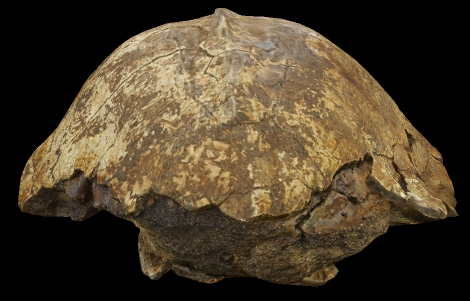
Homo naledi on my mind, and the lock screen on my phone, all year. CT rendering of cranium DH3, top is to the left and front is to the top.

I was in London for the ESHE conference last week when **it hit the fan, and so I got to attend a small press conference from the paper’s publisher, eLife, for the announcement.

eLife press conference last Thursday. From left to right: Will Harcourt-Smith, Matthew Skinner, Noel Cameron, Alia Gurtov and Tracy Kivell.

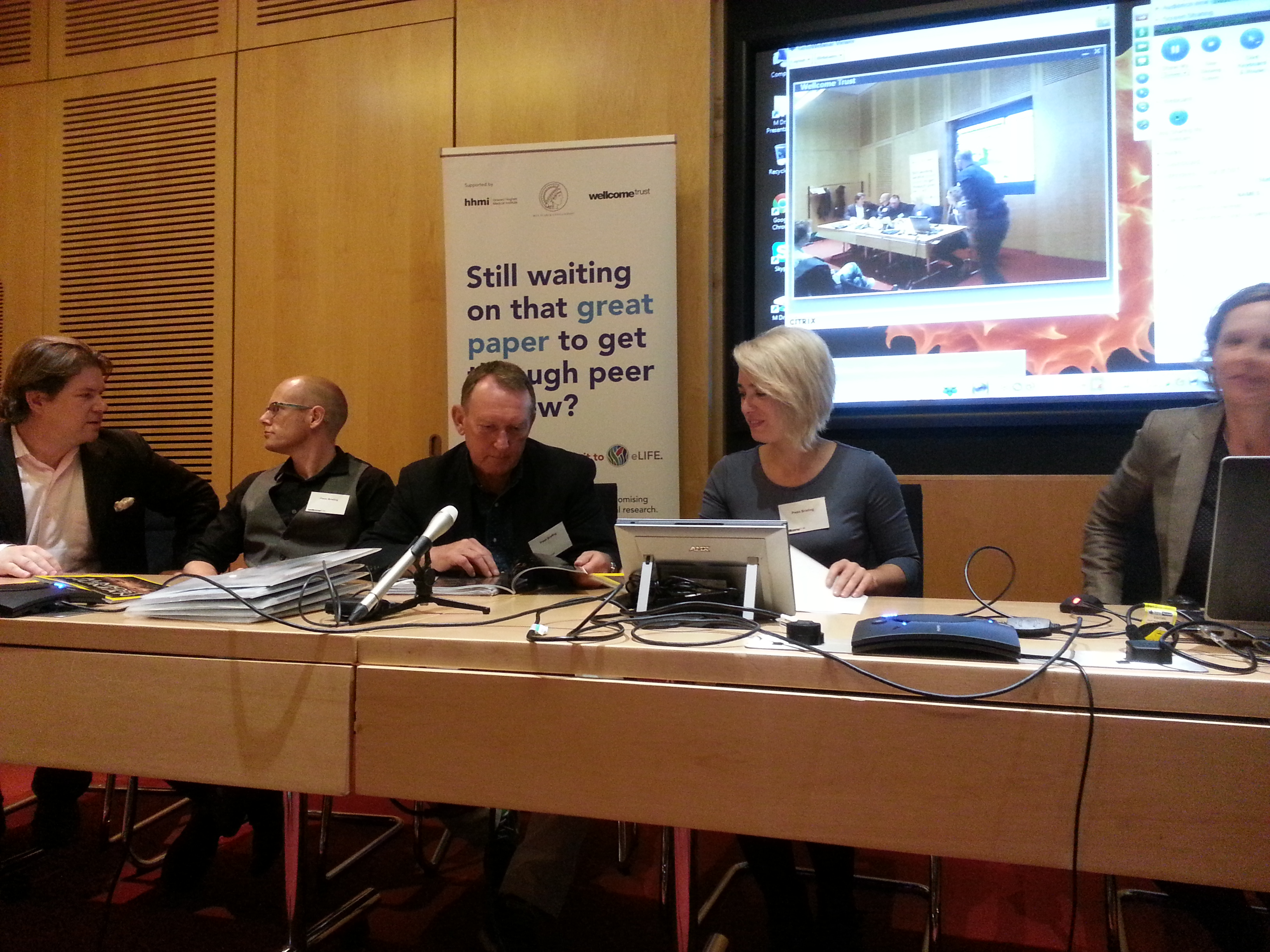
eLife press conference last Thursday. From left to right: friends and colleagues Will Harcourt-Smith, Matthew Skinner, Noel Cameron, Alia Gurtov and Tracy Kivell.

I had just flown in from Kazakhstan, and was presenting some recent work on the evolution of brain growth (I’ll write a post about it soon, promise), so it was a bit hard to appreciate the gravity of the announcement. Although the awesome spread in National Geographic did help it sink in a bit.

Really blurry photo of Markus Bastir holding up the heaviest copy of National Geographic ever.

I’m wending my way back to Kazakhstan now, but in the coming weeks I will try to post more about these fossils, the project, and specifically what I’m working on.

Until then, I’d like to point out how much information is freely and easily available to the entire world about these fossils. The paper, full-length and filled with excellent images of many of the specimens and reconstructions, is available for free online here. In addition, you can download 3D surface scans of over 80 of the original fossils on MorphoSource, also totally free. Everything about this scientific discovery and its dissemination is unprecedented – the sheer number of fossils and the ease of access with which literally everyone (well, with an internet connection) can access this information has never occurred before. This is the way paleoanthropology should be. Hats off to Lee Berger and the other senior scientists on the project for making such a monumental resource available to all.

ResearchBlogging.orgBerger LR, Hawks J, de Ruiter DJ, Churchill SE, Schmid P, Delezene LK, Kivell TL, Garvin HM, Williams SA, DeSilva JM, Skinner MM, Musiba CM, Cameron N, Holliday TW, Harcourt-Smith W, Ackermann RR, Bastir M, Bogin B, Bolter D, Brophy J, Cofran ZD, Congdon KA, Deane AS, Dembo M, Drapeau M, Elliott MC, Feuerriegel EM, Garcia-Martinez D, Green DJ, Gurtov A, Irish JD, Kruger A, Laird MF, Marchi D, Meyer MR, Nalla S, Negash EW, Orr CM, Radovcic D, Schroeder L, Scott JE, Throckmorton Z, Tocheri MW, VanSickle C, Walker CS, Wei P, & Zipfel B (2015). Homo naledi, a new species of the genus Homo from the Dinaledi Chamber, South Africa. eLife, 4 PMID: 26354291

Virtual paleontology activity

Last week Nazarbayev University hosted an Instructional Technology Showcase, in which professors demonstrated some of the ways we use technology in the classroom. This was the perfect venue to show off the sweet skeletal stuff we study in Biological Anthropology, through the use of pretty “virtual” fossils. In the past year I’ve started using CT and laser scans of skeletal remains to make lab activities in a few classes (I’ve posted two here and here). Such virtual specimens are especially useful since it is hard to get skeletal materials and casts of fossils here in the middle of the Steppe. These scans are pretty accurate, and what’s more, 3D printing technology has advanced such that physical copies of surface scans can be created from these virtual models. So for the Showcase, I had a table where passersby could try their hand at measuring fossils both in hand and in silico.

Lower jaw of an infant Australopithecus boisei (KNM ER 1477). Left is the plastic cast printed from the laser scan on the right.

Lower jaw of an infant Australopithecus boisei (KNM ER 1477). Left is the plastic cast printed from the laser scan on the right.

The Robotics Department over in the School of Science and Technology was kind enough to print out two fossils: KNM ER 1477, an infant Australopithecus boisei mandible, and KNM KP 271 a distal humerus of Australopithecus anamensis. They used a UP Plus 2 printer, a small desktop printer that basically stacks layers of melted plastic to create 3D models; they said it took about 9 hours to print the pair. Before the Showcase, I measured the computer and printed models on my own for comparison with published measurements taken on the original fossils (KP 271 from Patterson and Howells, 1967; ER 1477 from Wood, 1991). The virtual fossils were measured using the free program Meshlab, while basic sliding calipers were used to measure the printed casts.

I was pleasantly surprised at how similar my measurements were to the published values (usually within 0.1 mm), since it means that the free fossil scans provided by the National Museums of Kenya are useful not only for teaching, but potentially also for research.

The Virtual Paleontology Lab

The Virtual Paleontology Lab. The Kanapoi distal humerus is held in the foreground while the A. bosei jaw rests on the table. Yes, those are real palm trees.

Knowing that these models are pretty true to life (well, true to death, since they’re fossils), I was curious how students, faculty and staff would do. I picked two fairly simple measurements for each fossil. None of the people that came by to participate had any experience with bones or fossils, or measuring these in person or on a computer. Here are their results:

Boxplots showing participants' data, for two measurements on each of the fossils. The blue stars mark the published values. The red rugs on either side indicate measurements taken on the scans (left side) or printed casts (right).

Boxplots showing participants’ data, for two measurements on each of the fossils. The blue stars mark the published values. The red rugs on either side indicate measurements taken on the scans (left side) or printed casts (right).

For the most part, the inexperienced participants’ measurements are not too far off from the published values. There’s not really an apparent tendency for either cast or computer measurements to be more accurate, although measurements of the Kanapoi humerus are closer than the computer measurements (third and fourth boxes above). In my personal opinion, nothing beats handling fossils (or casts of them) directly, but this little activity suggests students can still make reliable observations using 3D scans on a computer.

Sweet free stuff:
Meshlab software
3D scans of fossils from the National Museums of Kenya

Calotte or Carapace?

Is this the top of a hominid skull, replete with sagittal crest running down the middle, or is it the top of a tortoise shell?

This image comes from great resource I just found (thanks to Louise Leakey on Twitter) for paleoanthropology students – africanfossils.org. I won’t answer here whether this is hominid or turtle, you’ll have to find it at the African Fossils site.

The site has 3D, manipulable images of fossil hominids and other animals from Kenya and Tanzania. The Smithsonian Museum of Natural History also has a very nice 3D collection, similarly manipulable. Resolution isn’t always what you might want it to be (for instance, you won’t be able to tell if the basi-occipital suture is fused in the Homo erectus cranium KNM-ER 42700), but you still get good overall view of some neat and bizarre animals. Like this robust australopithecus! (KNM-ER 406) Hey, its brain case does look kinda like the pic above…