Virtual paleontology activity

Last week Nazarbayev University hosted an Instructional Technology Showcase, in which professors demonstrated some of the ways we use technology in the classroom. This was the perfect venue to show off the sweet skeletal stuff we study in Biological Anthropology, through the use of pretty “virtual” fossils. In the past year I’ve started using CT and laser scans of skeletal remains to make lab activities in a few classes (I’ve posted two here and here). Such virtual specimens are especially useful since it is hard to get skeletal materials and casts of fossils here in the middle of the Steppe. These scans are pretty accurate, and what’s more, 3D printing technology has advanced such that physical copies of surface scans can be created from these virtual models. So for the Showcase, I had a table where passersby could try their hand at measuring fossils both in hand and in silico.

Lower jaw of an infant Australopithecus boisei (KNM ER 1477). Left is the plastic cast printed from the laser scan on the right.

Lower jaw of an infant Australopithecus boisei (KNM ER 1477). Left is the plastic cast printed from the laser scan on the right.

The Robotics Department over in the School of Science and Technology was kind enough to print out two fossils: KNM ER 1477, an infant Australopithecus boisei mandible, and KNM KP 271 a distal humerus of Australopithecus anamensis. They used a UP Plus 2 printer, a small desktop printer that basically stacks layers of melted plastic to create 3D models; they said it took about 9 hours to print the pair. Before the Showcase, I measured the computer and printed models on my own for comparison with published measurements taken on the original fossils (KP 271 from Patterson and Howells, 1967; ER 1477 from Wood, 1991). The virtual fossils were measured using the free program Meshlab, while basic sliding calipers were used to measure the printed casts.

I was pleasantly surprised at how similar my measurements were to the published values (usually within 0.1 mm), since it means that the free fossil scans provided by the National Museums of Kenya are useful not only for teaching, but potentially also for research.

The Virtual Paleontology Lab

The Virtual Paleontology Lab. The Kanapoi distal humerus is held in the foreground while the A. bosei jaw rests on the table. Yes, those are real palm trees.

Knowing that these models are pretty true to life (well, true to death, since they’re fossils), I was curious how students, faculty and staff would do. I picked two fairly simple measurements for each fossil. None of the people that came by to participate had any experience with bones or fossils, or measuring these in person or on a computer. Here are their results:

Boxplots showing participants' data, for two measurements on each of the fossils. The blue stars mark the published values. The red rugs on either side indicate measurements taken on the scans (left side) or printed casts (right).

Boxplots showing participants’ data, for two measurements on each of the fossils. The blue stars mark the published values. The red rugs on either side indicate measurements taken on the scans (left side) or printed casts (right).

For the most part, the inexperienced participants’ measurements are not too far off from the published values. There’s not really an apparent tendency for either cast or computer measurements to be more accurate, although measurements of the Kanapoi humerus are closer than the computer measurements (third and fourth boxes above). In my personal opinion, nothing beats handling fossils (or casts of them) directly, but this little activity suggests students can still make reliable observations using 3D scans on a computer.

Sweet free stuff:
Meshlab software
3D scans of fossils from the National Museums of Kenya

Australopithecus boisei bites

I always wondered what our extinct relative, Australopithecus boisei tasted like, until I moved to Kazakhstan.

2015-03-11 21.38.26

Mini calotte, or manti?

Here, dumplings with various fillings are called “manti” and usually have a distinct crimping running across the top. Along with their broad flaring bases and dome-like shapes, this gives manti the appearance of miniature A. boisei brain cases replete with sagittal crests:

They all look so delicious!

They all look so delicious! Fillings from left to right: lamb, pumpkin+lamb, mushrooms ewwwww.

In case you had trouble discerning braincase from блюдо, calotte from закуски in the pic, check out africanfossils.org and see if their handy, free 3D scans of fossils OH 5 and ER 406 help you figure it out.

eFfing #FossilFriday: Subfossil lemurs

Hard to resist the headline, “Enormous underwater fossil graveyard found,” from the National Science Foundation. The NSF posts a video detailing the discovery of an underwater cave system containing “hundreds of potentially 1,000-year-old [lemur] skeletons…” in Madagascar. As a paleontologist, hearing about the discovery large numbers of ancient skeletons is musical, like hearing Love This Giant or the new T Swift for the first time.

Two lemur crania in an underwater cave on Madagascar. Photo from nbcnews.com.

Two lemur crania in an underwater cave on Madagascar. Photo from nbcnews.com.

It’s a pretty remarkable discovery – hundreds if not thousands of bones representing many complete skeletons of various extinct lemur species. And toward the end of the clip is a skull of a pretty badass looking big cat. The video shows piles of loose bones dredged up from the cave. These will reveal lots of information about the biology of these recently extinct animals, especially if researchers can keep associated bones together.

So what are these animals? Lemurs are one of the most primitive living types of primates – although they are relatively closely related to us humans, they retain many characteristics of ancestral mammals. I know it’s hard to believe this aye-aye here is more closely related to you than to rodents, but it is:

An aye-aye (Daubentonia madagascarensis) using its narrow and elongated middle finger to fish for for grubs inside a tree that it’s opened up with its teeth.

Lemurs are found only on the island of Madagascar, and over the past several millions of years they have diversified into the roughly 100 species inhabiting the island today. But even just a few thousand years ago, there were more kinds of lemurs. This includes Megaladapis, the large-bodied “koala lemur,” and Hadropithecus, whose skull bears a striking resemblance to the extinct hominin Australopithecus boisei. As  Laurie Godfrey says in the video, “two thirds of the animals that lived there only a thousand years ago are gone.” Humans are probably largely responsible for the extinction of many Malagasy lemurs in both the past and especially the present.

Much of the ‘fossil’ record for lemurs is recent by fossil standards, and so most specimens haven’t become fully fossilized. As a result, lemur paleontology is besprinkled with the term “subfossil,” indicating bones that are really old and belong to extinct animals, but don’t fit the technical definition of fossils. The lemur subfossil record has taught us a lot about the evolutionary history, adaptations, and recently even genetics of this primitive group of primates, as well as about the ecological history of Madagascar. It will be very interesting to see what new insights will come from the recently discovered scores of underwater skeletons.

OH NO IT’S HADROPITHECUS

(Figure 3 from Ryan et al., 2008. Scale bar is 1 cm)

Another small Middle Pleistocene person

Last year I brought up the implications of the small female pelvis from Gona, Ethiopia for body size variation in Homo erectus (see previous post). This individual was much smaller than other Middle Pleistocene Homo fossils, indicating size variation comparable to highly sexually dimorphic gorillas and unlike recent human populations. Before this pelvis, most known Homo erectus fossils were fairly large (comparable to living people), with only a few hints of much smaller individuals (e.g., KNM-ER 427000, KNM-OL 45500). Now joining this petite party, this tiny troop, this little lot, this compact cadre, etc., is KNM-WT 51261, a 750,000 year old molar from Kenya (Maddux et al., in press).

Occlusal area for hominin first molars. The tooth is from Fig. 2 and the plot from Fig. 3 in the paper.

Occlusal area for first molars in the genus Homo. The tooth image is from Fig. 2 and the plot from Fig. 3 in Maddux et al. Lookit how tiny it is!

This ‘new’ specimen substantially increases the range of size variation among early African H. erectus molars, although the expanded range isn’t remarkable compared with later Homo samples such as from Zhoukoudian cave in China or Neandertals. What is different, though, is that most of the highly variable samples show a fairly continuous range of variation, while the WT 51261 molar is a considerable outlier from the rest of the African Middle Pleistocene sample (a lot like the situation with the Gona pelvis). So this tooth re-raises an important question: were smaller specimens like Gona and WT 51261 as rare in life as they are in the fossil record, or was such great size variation common in the Middle Pleistocene? How we reconstruct what kind of animal Homo erectus was differs depending on the answer to this question.

eFfing Fossil Friday: Funky facial flanges #FFF

David Krause and colleagues announced in this week’s Nature the discovery of a new species of extinct mammal, Vintana sertichi, that lived in what is now Madagascar between 66-72 million years ago. The species is based on a very well-preserved cranium of an early gondwanatherian (if you want to impress your friends this weekend, gratuitously use the word “gondwanatherian”). I don’t know much about early mammals like this, but it sounds like it was a weird creature (see the Stony Brook press release). Just looking at it’s face there’s something that sticks out as strange:

Ventana sertichi cranium (Reich et al. 2014, Figure 1a). Left is a 3D CT reconstruction, right is a line drawing highlighting all the individual bones (so many cranial bones). The view is from the right side, so the nose is on the right, the eye is the big hollow in the middle, and the back of the skull is on the left. The jugal flanges are the downward projections.

Vintana sertichi cranium (Reich et al. 2014, Figure 1a). On the left is a 3D CT reconstruction, and on the right is a line drawing highlighting all the individual bones (so many cranial bones). The view is from the right side, so the nose is to the right, the eye socket is the shadowy hollow in the middle, and the back of the skull is on the left. The jugal flanges are the downward projections.

Jutting downward from the sides of the jaw are ‘jugal flanges,’ projections of bone on the homologs of human cheeks. Projections of like these usually serve as muscle attachment sites, and the size of the projection generally reflects the size of the muscle. These facial flanges anchor the masseter muscle, a major chewing muscle that helps close the jaw. The size of this flange in Vintana suggests its chomp packed a punch. A debilitating bite. A face not even a mother could love (so now they’re extinct).

Vintana‘s bony tear-catchers caught my eye because most primates I’ve seen have, you know, less heinous faces. Scouring the internet, big jugal flanges are a fairly rare sight, but can apparently be found in glyptodonts (giant, armadillo-like mammals that lived tens of thousands of years ago) and various sloths. The closest thing I’ve seen to this gross bony flange in Primates are on the zygomatic bones of some extinct, baboon-like animals, such as Dinopithecus ingens:

Fragmentary skull, viewed from the top, of Papio (a.k.a. Dinopithecus) ingens, from Swartkrans, South Africa. Photo credit: CalPhotos.

Fragmentary skull, viewed from the top, of Papio (a.k.a. Dinopithecus) ingens, from Swartkrans, South Africa. As a punishment for its zygomatic excess, its face was confiscated. Photo credit: CalPhotos.

and Theropithecus brumpti

Theropithecus brumpti from the Omo basin. Photo credit: CalPhotos.

Theropithecus brumpti from the Omo Basin, Ethiopia. Photo credit: CalPhotos.

So some primates dabbled in jugal flangery like Vintana, but Natural Selection was having none of it. Anyway, Vintana overcame this craniofacial adversity with characteristic Mesozoic moxie, and is an important piece in the puzzle of mammal evolution. It will be interesting to see what other mammalian surprises the Mesozoic has in store for paleontologists.

eFfing Fossil Friday: resurrected

It’s been a quiet month here at Lawnchair, as I’ve just returned from the Rising Star Workshop, taking part in the analysis and description of new hominin remains from South Africa. We’ll have some exciting announcements to make in the near future.

Also, I petted a ferocious, bloodthirsty lion!20140601_160436

To ease back into the Lawnchair, I thought I’d resurrect eFfing Fossil Friday, a short-lived series from when I was collecting data for my dissertation three years ago (speaking of which, a paper related to my dissertation came out in AJPA during the Workshop, as well). A lot has happened since the last installment of FFF, so whose heads will be on the chopping block today?

Crania 9, 15 and 17 (clockwise from top left). Cranium 9 is an early adolescent and the other two are adults - lookit how the facial anatomy changes with age!

Crania #s 9, 15 and 17 (clockwise from top left). Cranium 9 is an early adolescent and the other two are adults – lookit how the facial anatomy changes with age! (Fig. 1 from Arsuaga et al., 2014)

It’s new crania from Sima de los Huesos, Atapuerca! These are published today in the journal Science by Juan L. Arsuaga and colleagues. Sima de los Huesos is a pretty remarkable site in Spain dating to the Middle Pleistocene; the site is probably at least 400,000 years old, and the remains of at least 28 individuals. These specimens show many similarities with Neandertals who later inhabited the area, but don’t have all of the ‘classic’ Neandertal features.

What I like about this figure from the paper is that the comparison of the adolescent (top left) with adults (the other two) shows how the skull changes during growth. The major visible difference is that the face sticks out in front of the brain case more in the adults than the adolescent. As a result, the adolescent lacks a supraorbital torus (“brow ridge”), but this would have developed as the face grew forward and away from the brain. Ontogeny!

Calotte or Carapace?

Is this the top of a hominin skull, replete with sagittal crest running down the middle, or is it the top of a tortoise shell?

This image comes from great resource I just found (thanks to Louise Leakey on Twitter) for paleoanthropology students – africanfossils.org. I won’t answer here whether this is hominin or turtle, you’ll have to find it at the African Fossils site.

The site has 3D, manipulable images of fossil hominins and other animals from Kenya and Tanzania. The Smithsonian Museum of Natural History also has a very nice 3D collection, similarly manipulable. Resolution isn’t always what you might want it to be (for instance, you won’t be able to tell if the basi-occipital suture is fused in the Homo erectus cranium KNM-ER 42700), but you still get good overall view of some neat and bizarre animals. Like this robust australopithecus! (KNM-ER 406) Hey, its brain case does look kinda like the pic above…

An end to Ediacaran embryology?

The things people can do these days. Therese Huldtgren and colleagues reported in last week’s Science that they identified nucleus-like structures in 570 million year old fossilized cells from China. These date to the Ediacaran period, before the “Cambrian explosion” of animal life forms. Superficially, these fossilized balls of cells rather resemble the early stages of animal embryos (see A in the figure below), in which cells are dividing and increasing in number but the overall embryo size stays the same. To get the “inside story” (…sorry), Huldtgren and team used very fancy “synchrotron x-ray computed tomography” to look at the insides of these fossilized cells. The resulting images have micrometer resolution – that’s one thousandth* of a millimeter. The things people can do these days.

Fig. 2 from Huldtgren et al. 2011

And lo! each of these fossilized cells contains a small, globular structure that looks like a nucleus (left; if you cross your eyes you can merge the 2 halves of fig. C to make it look even more 3D).

Could these really be the earliest animal embryos? Probably not – some of these balls-of-cells had what resemble budding spores, unlike animals but similar to “nonmetazoan [non-animal] holozoans.” In other words, something neat and old, but not one of our earliest ancestors.

I’m really impressed with the biological applications of computed tomography (CT). Recall that a while ago, I posted about the potential to use synchrotron tomography to examine the small-scale, internal structure of bone (e.g. Cooper et al. 2011). Such non-destructive, high-resolution imaging techniques could be used to compare near-cellular-level growth in living and fossil animals. This is really significant because it adds a completely new kind of information we can get from fossils, which before now could only be studied well at the gross, macroscopic level (though scanning electron microscopy of teeth has been very informative about diet; see for example Ungar and Sponheimer 2011). Indeed, one of the most common applications of CT imaging in anthropology is making 3D computer models of body parts for morphometric (shape) analysis.

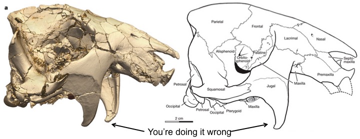
But high-resolution, synchrotron CT imaging opens up a whole new world of paleontology, new questions that can be asked. For example, many researchers have examined the microscopic appearance of bone surfaces to determine whether bone was being added or removed during growth, and comparing different species (Bromage 1989, O’Higgins et al. 2001, McCollum 2008, Martinez-Mata et al. 2010). These have been very informative studies, but it is not totally clear how growth at the cellular level relates to growth at visible level. Moreover, fossil surfaces are often abraded, obfuscating surface details. So, I can envision using synchrotron microscopy similar to Cooper et al. (2011) and Huldtgren et al. (2011), to examine bone growth in fossil hominids, at and beneath the surface. This can help us understand how facial growth was modified over the course of human evolution, from the snouty visage of Australopithecus afarensis to the tiny, starry-eyed faces we have today. People could also examine how activities like chewing, running or even talking affect (and effect) bone growth. There is much work to be done.

ResearchBlogging.orgNeat as these projects would be, it’s pretty humbling to consider that we have the technology to analyze microscopic fossils hundreds of millions of years old, and shed light on the developmental processes in our earliest ancestors.

Read those things I’d mentioned

BROMAGE, T. (1989). Ontogeny of the early hominid face Journal of Human Evolution, 18 (8), 751-773 DOI: 10.1016/0047-2484(89)90088-2

Cooper, D., Erickson, B., Peele, A., Hannah, K., Thomas, C., & Clement, J. (2011). Visualization of 3D osteon morphology by synchrotron radiation micro-CT Journal of Anatomy, 219 (4), 481-489 DOI: 10.1111/j.1469-7580.2011.01398.x

Huldtgren, T., Cunningham, J., Yin, C., Stampanoni, M., Marone, F., Donoghue, P., & Bengtson, S. (2011). Fossilized Nuclei and Germination Structures Identify Ediacaran “Animal Embryos” as Encysting Protists Science, 334 (6063), 1696-1699 DOI: 10.1126/science.1209537

Martinez-Maza, C., Rosas, A., & Nieto-Diaz, M. (2010). Brief communication: Identification of bone formation and resorption surfaces by reflected light microscopy American Journal of Physical Anthropology, 143 (2), 313-320 DOI: 10.1002/ajpa.21352

McCollum, M. (2008). Nasomaxillary remodeling and facial form in robust Australopithecus: a reassessment Journal of Human Evolution, 54 (1), 2-14 DOI: 10.1016/j.jhevol.2007.05.013

O’Higgins, P., Chadfield, P., & Jones, N. (2001). Facial growth and the ontogeny of morphological variation within and between the primates Cebus apella and Cercocebus torquatus Journal of Zoology, 254 (3), 337-357 DOI: 10.1017/S095283690100084X

Ungar, P., & Sponheimer, M. (2011). The Diets of Early Hominins Science, 334 (6053), 190-193 DOI: 10.1126/science.1207701

eFfing Fossil Friday (another late edition)

ResearchBlogging.orgI’m sitting at a cafe in Tbilisi, departing at 4:00 am tomorrow for America. Readers will notice that I’ve been MIA while working with the second annual Dmanisi Paleoanthropology Field School. I hate to say it but I’m glad I was too busy to blog all the goings-on (though sorry if it disappointed anyone). All in all it was another great year, and we found some great fossils (about which I don’t think I have permission to say anything at all). Here’s this year’s class with their certification of badassery at the site on the last day:
But Dmanisi won’t be the subject of this belated eFfing Fossil Friday. I’d like instead to turn to the question of just what fossils are good for. I’m told that in China, fossil teeth were once interpreted as dragons’ teeth, and so pulverized and sold as medicine. But what good are they to non-medical science? My recent research interests have come to focus on the relationship between evolution and development. Evolutionary developmental biology (“evo-devo”) research has been dominated by studies of genes, gene expression, and model organisms like fruit flies and mice. In such an environment, the question of the relevance of fossils is especially poignant.
But this morning, while planning a human evo-devo course I hope to teach next summer, I stumbled upon a review paper by Rudolf Raff, titled “Written in Stone: Fossils, genes and evo-devo” (2007). I think the abstract sums things up pretty well:

Fossils give evo-devo a past. They inform phylogenetic trees to show the direction of evolution of developmental features, and they can reveal ancient body plans. Fossils also provide the primary data that are used to date past events, including divergence times needed to estimate molecular clocks, which provide rates of developmental evolution. Fossils can set boundaries for hypotheses that are generated from living developmental systems, and for predictions of ancestral development and morphologies. Finally, although fossils rarely yield data on developmental processes directly, informative examples occur of extraordinary preservation of soft body parts, embryos and genomic information.

It seems often that fossils are falling by the wayside. There’s a sentiment that there’s not much information to be gotten from fossils – they’re too incomplete, too few, too inconvenient, at least as compared with extremely high-output data such as that coming from genomics. But Raff is right – we need fossils. Beyond the excellent points Raff raises in the review, I’m working on getting the most out of these seemingly data-poor fossil samples. Because modern computers are so powerful nowadays, I’m using their sheer processing power to test hypotheses about growth and development in fossil samples. These battered bunches of bones are too tiny to be analyzed by traditional methods. But one thing I think is important to take away from this computer-crazy Information Age, is that we now have machines that can handle almost any kind of question one can think to ask, and it’s really inspiring. The sequencing and analyses of ancient Neandertal and Denisova genomes (Green et al. 2010, Reich et al. 2010) are excellent examples of the amazing research that can be done with computers and creativity (and probably also a horde of hard-working math majors).
So this eFFF (or Sunday) is not dedicated to any specific fossil or set of fossils, but rather to all fossils, even the crappy fragments. Gaumarjos, fossils: your secrets are not safe from us.
Reference
Green, R., Krause, J., Briggs, A., Maricic, T., Stenzel, U., Kircher, M., Patterson, N., Li, H., Zhai, W., Fritz, M., Hansen, N., Durand, E., Malaspinas, A., Jensen, J., Marques-Bonet, T., Alkan, C., Prufer, K., Meyer, M., Burbano, H., Good, J., Schultz, R., Aximu-Petri, A., Butthof, A., Hober, B., Hoffner, B., Siegemund, M., Weihmann, A., Nusbaum, C., Lander, E., Russ, C., Novod, N., Affourtit, J., Egholm, M., Verna, C., Rudan, P., Brajkovic, D., Kucan, Z., Gusic, I., Doronichev, V., Golovanova, L., Lalueza-Fox, C., de la Rasilla, M., Fortea, J., Rosas, A., Schmitz, R., Johnson, P., Eichler, E., Falush, D., Birney, E., Mullikin, J., Slatkin, M., Nielsen, R., Kelso, J., Lachmann, M., Reich, D., & Paabo, S. (2010). A Draft Sequence of the Neandertal Genome Science, 328 (5979), 710-722 DOI: 10.1126/science.1188021


Raff, R. (2007). Written in stone: fossils, genes and evo–devo Nature Reviews Genetics, 8 (12), 911-920 DOI: 10.1038/nrg2225
Reich D, Green RE, Kircher M, Krause J, Patterson N, Durand EY, Viola B, Briggs AW, Stenzel U, Johnson PL, Maricic T, Good JM, Marques-Bonet T, Alkan C, Fu Q, Mallick S, Li H, Meyer M, Eichler EE, Stoneking M, Richards M, Talamo S, Shunkov MV, Derevianko AP, Hublin JJ, Kelso J, Slatkin M, & Pääbo S (2010). Genetic history of an archaic hominin group from Denisova Cave in Siberia. Nature, 468 (7327), 1053-60 PMID: 21179161

eFFING FOSSIL FRIDAYS!

I’m going to do my best to keep up with the blog during by Big Summer Adventure, and one thing I’d like to do is “F-ing Fossil Friday!” in which I focus on fossils for a bit. We’ll see if I can make this pan out.
Today I got out the rest of the Australopithecus robustus mandibles at the Transvaal Museum (above), save for I think maybe 1. As you can see from the picture, taphonomy (what happens to an animal’s remains between death and our digging them up) creates a serious challenge for the study of variation in this species. I’m focusing on ontogenetic variation – differences associated with growth and development. In spite of its fragmentary nature, so far as I know this is the best ontogenetic series of any fossil hominid (I should probably look more into A. afarensis here, too). In the bottom left you’ll see SK 438, the youngest in the sample, whose baby teeth haven’t quite come in all the way. Poor little guy! At the top right corner is SK 12, probably the oldest individual and also a big bugger.
One thing that I’ve noticed so far, only a preliminary observation that I need to actually run some numbers on, is that as individuals get older, the length of their tooth row (molars and premolars) gets shorter. This is because of the tendency for teeth to move forward during growth – “mesial drift” – and for adjacent teeth to literally wear into one another, their ends becoming flatter and flatter. While I should have realized this, it was surprising at first to find some dimensions of the lower jaw actually decreasing during growth. Now, I still have to run some tests to see if this is a biologically significant phenomenon. But it’s always nice to learn something new, even after just 2 days back with my best extinct buddies.
Stay tuned to future eFfing fossil Fridays!