Climate change works fast

A study just came out in Science showing that the water cycle – the process of water being evaporated to the atmosphere, condensed into clouds, and returned to Earth as rain – has sped up dramatically in just the past 50 years (Durack et al. 2012). From news coverage of the research (Kerr 2012), here’s a reason why this speed-up sucks and has the potential to suck more:

Such a revved-up water cycle would have “a lot of implications for how extreme events would change in a warming climate,” says meteorologist Brian Soden of the University of Miami in Florida. Water cycling from the surface to the atmosphere carries heat energy that can ultimately fuel violent storms, from tornadoes to tropical cyclones. The faster water cycles, the more abundant and more violent those storms might be. And wet places getting wetter can lead to more severe and more frequent flooding. Dry places getting drier would mean longer and more intense droughts.

Durack and colleagues’ findings are important because they show just how rapidly and drastically the Earth is changing, right before our eyes. Unlike humans, most plants and animals are adapted to fairly specific ecological circumstances, and departure from the norm can spell extinction, especially in long-lived, slow-reproducing species. We humans are adept at altering our environment to our likings, and until recently we’ve managed to avoid (or at least be ignorant of) the consequences of our earthworks. This is serious stuff that we can actually do something about, but only if we make scientifically-informed decisions.

ResearchBlogging.orgI don’t know that I’ve ever gotten political on this blog, but I’d like to stress now that climate change is an issue people should be thinking about in this election year. The Republican primaries have largely been centered around shitshow discussions of straw man issues and Dominionist fluff – it would have been laughable if none of those clowns were seriously trying to become the president. But now that Romney will be the Republican candidate to run against Obama, hopefully debates will come down to real world issues. (Read more about the role of climate change in candidates’ campaigns here at the Huffington Post)

UPDATE 02 SEPTEMBER – Nope. The Republican National Convention was just held in Tampa, FL, where Mitt Romney mocked Obama’s pledge to address climate change (not as bad as critics made him sound). Mitt and his running mate Paul Ryan have insisted for the past few weeks that they’d bring solutions to the issues they say Democrats have failed to address, yet these Republican candidates have continuously lied about the past and been deplorably vague about how they plan to improve America. It will be upsetting on several levels if these buffoons end up in the White House.

The good news & the bad news
Durack, P., Wijffels, S., & Matear, R. (2012). Ocean Salinities Reveal Strong Global Water Cycle Intensification During 1950 to 2000 Science, 336 (6080), 455-458 DOI: 10.1126/science.1212222

Kerr, R. (2012). The Greenhouse Is Making the Water-Poor Even Poorer Science, 336 (6080), 405-405 DOI: 10.1126/science.336.6080.405

An unusual hominin foot

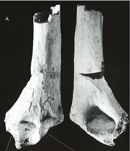
Researchers announced in Nature today the discovery of a 3.4 million-year-old foot that doesn’t “toe the hominin line.” Dammit I regret that already. Anyway, Ethiopian paleoanthropologist Yohannes Haile-Selassie and colleagues have found the foot of a creature whose big toe was oriented away from the rest of the foot and capable of grasping, like all primates (including Ardipithecus ramidusexcept later hominins. See for yourself:

BRT-VP-2/73 foot bones. Look at that fat, abducted hallux! And too-long 4th metatarsal! (fig. 1 from the paper)
World’s greatest left foot.

To help you orient yourself, the left third of the above figure (labeled with a tiny “a”) is a top-view of the ‘articulated’ right foot of this mystery animal. To the right is an X-ray (or “roentgenogram,” if you’re so inclined) of my left foot.

This is an immensely exciting find. The fossils are from a site in Ethiopia called Burtele dating to around 3.4 million years old. This is 1 million years after Ardipithecus ramidus from Aramis (also in Ethiopia), and contemporaneous with Australopithecus afarensis (also Ethiopian, viz. sites like Maka, Dikika and the earlier parts of the Hadar formation). With its divergent, grasping big toe, we can be pretty sure this foot did not belong to Au. afarensis, the maker of the famous Laetoli Footprints which are a few hundred thousand years older than the Burtele foot. Other aspects of the foot, however, like the round, “domed” heads of the metatarsals and the upward-angling of the proximal toe-bones do suggest this thing may have been bipedal in light of its grasping big toe (or shall we say, “foot-thumb”). Now, this upward canting of proximal toe bones’ proximal ends is associated with bipedality, specifically hyper-dorsiflexing (or hyperextension) of the toes – this movement doesn’t necessarily have to come solely during bipedalism, and we have some baboon proximal toe bones in our lab that have slight angling (admittedly, though, not as strongly as in humans).

From the metric analyses of the foot, a few major things stick out. First, where the Burtele foot is similar to humans, both species are also extremely similar to gorillas. The plots at right, from the paper, show the height of the first metatarsal’s (foot-thumb’s) base relative to its length (a), and relative to the base height of the second metatarsal (b). The first plot shows that, compared with chimpanzees and Old World monkeys, the foot-thumb’s base is fairly tall relative to its length. Here, the fossil is smack within the highly-overlapping human and gorilla ranges. The second plot shows that, compared with monkeys, all apes (including humans) and the fossil have tall first metatarsal bases relative to the height of the second metatarsal. Notice that the human and gorilla ranges overlap, though humans are a little higher; here the fossil is at the far end of the human range with a very tall foot-thumb base. Finally, in a principal components analysis of foot bone ratios, humans and gorillas overlap a bit, to the exclusion of chimpanzees and monkeys, and the fossil plots within the gorilla (but not human) range. What really gets me here is the remarkable similarity between humans and gorillas. Since metric analyses indicate that the gorilla-human similarities are largely confined to the aspects foot-thumb, I’d imagine the similarity is due to (1) humans’ putting greater force on our big toes because we walk on two legs, and (2) gorillas’ putting lots of force on their foot-thumbs because they are massive, massive animals. Either way, the Burtele foot-thumb is so similar to both of us. Another interesting thing revealed by Haile-Selassie et al.’s analyses is that Burtele’s fourth metatarsal is extremely long, unlike African apes (including humans), but more similar to cercopithecine monkeys and the 20 million-year-old early ape Proconsul. The authors take this to suggest that a long 4th metatarsal is the primitive condition for apes.

And so what I think is so exciting and important about the Burtele fossils is that they further demonstrate that we have a ton to learn about human our past that only the fossil record can illuminate (the recent Ardipithecus ramidus, Australopithecus sediba and the Woranso-Mille A. afarensis skeletons highlight this as well). The authors say the Burtele fossils demonstrate a second kind of bipedalism in a hominin lineage separate from the contemporaneous A. afarensis. But since we have no idea what the rest of this animal looked like, it raises the intriguing possibility that we may finally have a fossil ancestor  or relative of the other apes. I’ve long been suspicious that nearly every single ape/human-like fossil found in Africa younger than 7 million years is attributed to the hominin line, so I’d be very pleased if this turned out to be a non-hominin ape (though again I don’t necessarily think that’s what the Burtele fossils are).

The paper itself:

Haile-Selassie, Y., Saylor, B., Deino, A., Levin, N., Alene, M., & Latimer, B. (2012). A new hominin foot from Ethiopia shows multiple Pliocene bipedal adaptations Nature, 483 (7391), 565-569 DOI: 10.1038/nature10922

Avoid the Noid… I mean Noise

As alluded to yesterday, my dissertation compares growth in an extinct animal with growth in living humans; this study is necessarily cross-sectional, meaning that it examines individuals at a single point in time. Alternatively, longitudinal data sample individuals from several points in time. So for instance if I constructed a growth curve by measuring the stature of a bunch of people of different ages in just a day, that would be cross-sectional. But if I had the time and wherewithal to measure some people’s heights once a year from birth to adulthood, well that’d be longitudinal. Cross-sectional data lack the resolution of longitudinal data, whereas longitudinal data can be prohibitively difficult to collect (such as in long-lived, slow-maturing animals like humans, or in extinct animals like Australopithecus robustus).

Some researchers abhor cross-sectional data, pointing out that the intricacies of individuals’ longitudinal growth will not be adequately captured in with cross-sectionally. American anthropology founder Franz Boas himself discussed this in a paper nearly 82 years ago. Anyway, I was reminded of this dichotomy today when perusing a paper that examined longitudinal brain activity in a cohort of adolescent kids (right, from Campbell et al. in press). The mess of jagged lines are individuals’ measurements from age 9-18, and the smoothed blue and red curves are the cross-sectionalized curves calculated from these kids. Oy, look at all that variation and caprice that gets left out in the cross-sectionalized curves!

Of course, this doesn’t mean that we should never use cross-sectional data to study growth – like I’d mentioned above, the fossil record necessitates a cross-sectional approach to the study of growth. As always, you have to understand and acknowledge the limits of your data.

ResearchBlogging.orgRead on
Boas, F. (1930). OBSERVATIONS ON THE GROWTH OF CHILDREN Science, 72 (1854), 44-48 DOI: 10.1126/science.72.1854.44

Campbell, I., Grimm, K., de Bie, E., & Feinberg, I. (2012). Sex, puberty, and the timing of sleep EEG measured adolescent brain maturation Proceedings of the National Academy of Sciences DOI: 10.1073/pnas.1120860109

It’s quiet…

Although things have been silent here at Lawnchair Anthropology, this isn’t because there’s nothing going on. Rather, there’s a tonne going on right now and I haven’t had the time to write here. A few weeks ago I accepted a job, starting in August, in the burgeoning anthropology department of Nazarbayev University in Kazakhstan. It’s a brand new school and I’ll be the first biological anthropologist there (I believe), which is exciting. But, this means I need to book it to finish my dissertation by August. Also I’m teaching a Human Evo-Devo class (Anthrbio 297) here at the University of Michigan in the Spring (May-June) which will be awesome but will definitely make it challenging to get this thesis finished. I’ll do it, though.

So things’ve been pretty crazy lately, and I don’t foresee that changing any time soon. I’ll do my best to keep things up to date here at Lawnchair. The annual meetings of the American Association of Physical Anthropologists are happening in Portland in a few weeks, so I’ll probably have stuff to report on from there (if not here on lawnchair, then at least as soundbites on Twitter).

Osteology everywhere: Pelvis has left the building

The vernal awakening has brought rain to Ann Arbor, and right on here on main campus I spotted the rain-splotched silhouette of an articulated human pelvis (left).

Check out those short and flaring iliac blades, and the shortness of the ischium. These features are associated with repositioning key muscles for walking and running on two feet, and are very unlike what is seen in the four-legged, suspensory climbing apes.

Calotte or Carapace?

Is this the top of a hominin skull, replete with sagittal crest running down the middle, or is it the top of a tortoise shell?

This image comes from great resource I just found (thanks to Louise Leakey on Twitter) for paleoanthropology students – africanfossils.org. I won’t answer here whether this is hominin or turtle, you’ll have to find it at the African Fossils site.

The site has 3D, manipulable images of fossil hominins and other animals from Kenya and Tanzania. The Smithsonian Museum of Natural History also has a very nice 3D collection, similarly manipulable. Resolution isn’t always what you might want it to be (for instance, you won’t be able to tell if the basi-occipital suture is fused in the Homo erectus cranium KNM-ER 42700), but you still get good overall view of some neat and bizarre animals. Like this robust australopithecus! (KNM-ER 406) Hey, its brain case does look kinda like the pic above…

You may have my statistical codes

As I’ve been working on my dissertation, I’ve had to come up with some new ways to compare (cross-sectional) growth in crappy fossil samples with a larger reference population. I’ve coded a procedure in the R statistical program that uses resampling to test whether two groups differ in the amount of size change experienced between various different ages (i.e. growth). This code is now available on my website.**

And how timely – a commentary in this week’s issue of Nature demands that researchers publish the codes used in their analyses (Ince et al. 2012). After all, what good is Science if it’s not reproducible? (Admittedly, the commentary is geared toward more intense, data-generating programs than anything I’ve written, which is mathematically very simple and generally comprises less than 100 lines of code. Nevertheless.)

ResearchBlogging.orgAnyone is free to use or adapt the code, with the caveat that one must have at least a little experience using R. In many ways the procedure is similar to a method called Euclidean Distance Matrix Analysis (EDMA; Lele and Richtsmeier 1991), although unlike EDMA my program centers around the problem of making comparisons in the face of lots of missing data. And lots of fun!

**  Oh crap! I just remembered I also posted a simple resampling procedure here on Lawnchair two and a half years ago. Where does the time go…

Some inspiration
Ince, D., Hatton, L., & Graham-Cumming, J. (2012). The case for open computer programs Nature, 482 (7386), 485-488 DOI: 10.1038/nature10836

Lele, S., & Richtsmeier, J. (1991). Euclidean distance matrix analysis: A coordinate-free approach for comparing biological shapes using landmark data American Journal of Physical Anthropology, 86 (3), 415-427 DOI: 10.1002/ajpa.1330860307

Osteology Everywhere

I saw a humerus bone sticking out of the ground on my walk home today.

Just kidding. It was just a stupid tree (left). But it does look a lot like a reversed back-side view of thASK-VP-3/78 distal humerus of Ardipithecus kadabba (right-most of the right pic; Haile-Selassie 2001). It’s like someone blew up and unacceptably interred it, exposing only the top of the olecranon fossa (the big pit in the pic on the right, where the roots bifurcate on the tree at left). “ARE YOU A HOMINID OR NOT?” I almost yelled at the tree.
When you spend so much of your time working with bones, well you start seeing bones everywhere. And you’d be surprised how often you’ll find something when you’re looking for it, even inadvertently.
ResearchBlogging.orgWhat nature reminded me of: Haile-Selassie Y (2001). Late Miocene hominids from the Middle Awash, Ethiopia. Nature, 412 (6843), 178-81 PMID: 11449272

Ameloblast from the past

I’ve posted a couple times about the prospects of using high-resolution computed tomography imaging to assess cellular-level processes of growth and development. Today, Paul Tafforeau and colleagues present a synchrotron-based visualization of the adventurous paths that individual enamel-forming cells'(ameloblasts) take to form tooth crowns. I’ve been focusing more on using these techniques for studying bone growth, but I got the idea of that from previous studies of teeth (see Macchiarelli et al. 2006 and Smith et al. 2010).

Tafforeau et al 2012, Fig 3. Scale bar = 0.25 mm

Time was, the internal microstructure and growth of enamel could only be examined using sectioned (either cut or naturally fractured) tooth crowns. Synchrotron imaging of teeth allowed Tafforeau and colleagues to get at this internal information in complete teeth whose insides are unexposed.

To the left is a “virtual” section of a molar tooth, the ‘base’ of the enamel (at the enamal-dentine junction) is at the bottom right, and the external surface of the tooth is at the top left. The lines radiating from the EDJ to the crown surface are enamel prisms, the mineralized paths of cells called “ameloblasts” that form tooth crowns. This is the cellular process by enamel is deposited to form a rock-hard tooth.

Note that the prisms start off packed closely together as they start their journey from the EDJ, but slowly diverge along roughly-parallel paths to be a bit further apart from one another (cross-sections in the cubes). The prisms’ shadow on projected onto the exposed crown shows how non-linearly ameloblasts course to their final destination in some dimensions – I for one don’t know why the path contains these kinks.

As with any awesome method, there are nevertheless limitations. Tafforeau and team say that enamel closer to the inside of the tooth is somewhat muddled, due to differences in the extent to which prisms had mineralized. And I don’t know any numbers, but I’d guess that scanning a lot of teeth would get pretty expensive. But ultimately is a pretty badass research tool. This fine-scale internal view of tooth microstructure can allow researchers to reconstruct how a tooth grew, and from there to examine the cellular growth processes involved in certain crown shapes, mechanical properties of teeth, and how enamel hypoplasias (markers of health stress) are created by affecting the behavior of cells. Very cool stuff.

ResearchBlogging.orgThose papers
Macchiarelli, R., Bondioli, L., Debénath, A., Mazurier, A., Tournepiche, J., Birch, W., & Dean, M. (2006). How Neanderthal molar teeth grew Nature, 444 (7120), 748-751 DOI: 10.1038/nature05314

Smith, T., Tafforeau, P., Reid, D., Pouech, J., Lazzari, V., Zermeno, J., Guatelli-Steinberg, D., Olejniczak, A., Hoffman, A., Radovcic, J., Makaremi, M., Toussaint, M., Stringer, C., & Hublin, J. (2010). Dental evidence for ontogenetic differences between modern humans and Neanderthals Proceedings of the National Academy of Sciences, 107 (49), 20923-20928 DOI: 10.1073/pnas.1010906107

Tafforeau, P., Zermeno, J., & Smith, T. (2012). Tracking cellular-level enamel growth and structure in 4D with synchrotron imaging Journal of Human Evolution DOI: 10.1016/j.jhevol.2012.01.001